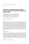
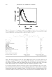
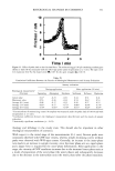
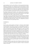
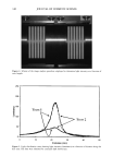
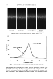
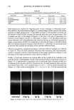
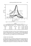
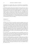
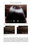
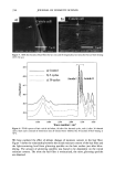
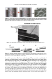
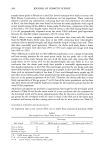
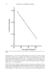
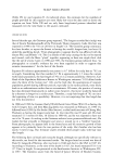
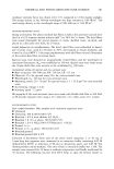
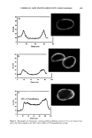
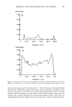
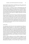
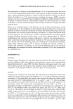
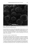
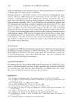
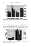
402 JOURNAL OF COSMETIC SCIENCE 25 2O 15 10 Hair ß Sepia 0 20 40 60 8O 100 Time (min) Figure 4. Time-dependent fluorescence study for the bleaching of hair and Sepia melanin using NH3/H202 at pH 10.0. A suspension of melanosomes (0.5 mg/ml) was treated with 2% H202 and 1.0% ammonia, and aliquots were taken every 5 min for time-dependent measurements. Intensity of fluorescence emission is plotted as a function of time. hydroxide or sodium carbonate treatment at pH 10 does not effect similar structural changes to hair melanosomes as ammonia. Thus, it is confirmed that the observed melanosomal changes are not merely due to alkaline pH but are, rather, specific to ammonia. Sepia melanosomes under identical conditions of ammonia treatment did not show such changes, probably due to a slow rate of bleaching, as evident from the time-dependent fluorescence studies. Prolonged treatment of hair melanosomes with NH3 induced a complete destruction of characteristic melanosome morphology and a shrinking of the granule size (Figure 8). As shown in Figure 8, the melanosomes appeared as fibril-type structures devoid of the melanosomal membrane sac. After 3 hr of treatment, no particles were seen by TEM, except an amorphous material. The ammonia-treated melanin suspension showed a lighter color compared to the untreated suspension, and a decrease in longer wavelength absorbance in the UV-Vis spectrum. Thus, it is evident that ammonia could possibly induce lightening of hair melanosomes. However, the overall process of particle break- down leading to an amorphous material is slow and took more than 3 hr. TEM STUDIES ON THE EFFECT OF HYDROGEN PEROXIDE Isolated hair melanosome suspension was treated with 2% n20 2 solution at pH 7.0 and pH 8.0 to see how peroxide alone degrades melanosomes. The degradation of melano- somes by n202 at neutral pH is a very slow process, and even after 60 min of treatment,

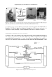
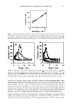
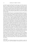
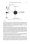
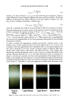
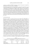
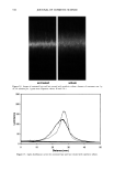
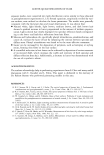
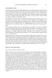
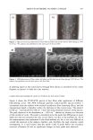
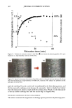
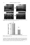
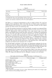
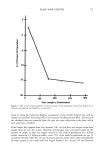
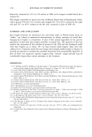
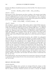
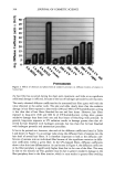
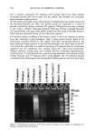
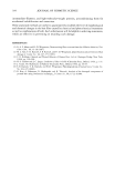
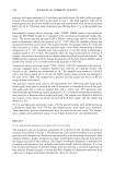
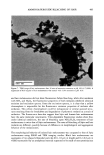
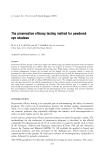
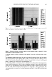
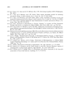
AMMONIA/PEROXIDE BLEACHING OF HAIR 403 Figure 5. ESEM image of a suspension of Sepia melanin (x4000). A suspension of 0.05 mg/ml of Sepia melanosomes was used for the ESEM imaging studies. intact melanosomes were visible by TEM image analysis. However, after 2-3 hr of treatment, structural changes leading to the dissolution of melanin granules were ob- served. The changes were slightly faster at pH 8.0, and prolonged treatment led to a clear solution, indicating that bleaching has occurred. The study clearly suggests that neutral H202 is less efficient than NH 3 in inducing morphological and structural changes to hair melanosmes. TEM STUDIES ON THE EFFECT OF AMMONIA/PEROXIDE Time-dependent TEM imaging studies were performed on isolated hair melanosomes (0.05 mg/ml) after treatment with NH 3 (1%) and H202 (1%) at pH 10. Co-treatment with NH 3 and H202 induced a faster disintegration of melanosome particles, and structural changes were visible even after 10 min of treatment. Figure 9 shows the TEM image of ammonia/peroxide-treated hair melanosomes after 15 min of treatment. In contrast to the effect of ammonia, co-treatment with ammonia and peroxide induced a simultaneous disintegration and dissolution of melanosomes. The overall disintegra- tion, dissolution, and discoloration process is much faster than ammonia treatment due
Purchased for the exclusive use of nofirst nolast (unknown)
From: SCC Media Library & Resource Center (library.scconline.org)