204 JOURNAL OF COSMETIC SCIENCE accepted that the secondary structure and property of fibrous proteins such as hair keratins, which are major components in the hair cortex and which stabilize their quaternary structure by a disulfide-bridge, are correlated with their strength by enhanc ing covalent cross-links between polypeptide chains within the multihelical ropes and between adjacent chains in a supramolecular assembly. Accordingly, the biochemical basis of permanent waving is derived from the characteristic stretchability of a-keratins, which is caused by a change in a-helical conformation (3). In general, some factors and chemical properties of the waving lotion used in the process of permanent waving treatment affect the hair change. For example, the permanent waving lotions containing the reactive thiol or sulfhydryl groups (-SH) have been used to reduce the disulfide bonds of the hair and to form a permanent wave by allowing molecular changes in its component proteins (1,2,4-6). In addition, other factors such as heat and pH influence the permanent wave (1,2). Furthermore, repeated permanent waving treatments also influence the hair. The factors and the chemical properties described above play important roles in the process of permanent waving. Some workers reported that permanent treatment induced changes in some chemical compositions of hair such as a decrease in cystine and cysteic acid (2), elution of certain proteins (7) and changes in the a-helix content (6,8). Other research groups have provided some evidence that physicomorphological properties, such as changes in tensile strength and hair shape, hair elongation, diameter, and degree of swelling, were altered by the perming treat ments (2,9). Other workers conducted light and electron microscopic investigations and demonstrated various characteristic changes in the surface of human hairs after perma nent waving treatment (1,9-11). These microscopic observations showed that change on the surface of the hair shaft and the degree of hair damage occurring from treatment with the chemical solutions or during the permanent waving process depend on the perming method (1,2). The above studies are informative for human hair care because they can be utilized for minimizing hair damage arising from perming treatment. Consequently, this study is aimed at presenting some information about changes in protein content and profile in the hair shafts of Korean women, performed with two methods of permanent waving: the croquignole winding perm (CWP, a type of normal perm characterized by wrapping hair from the ends to the scalp with no thermal conditioning) and the digital perm (DP, a perm processed by a special perm machine to give a thermal-conditioning permanent wave). These methods are more popular than others for most Korean women. In addition, some changes in the physical properties of the hair shafts and their mor phological images observed with a scanning electron microscope are also reported and discussed in relation to the perming treatment. MATERIALS AND METHODS HUMAN HEAD HAIR MATERIALS For protein extraction from human head hairs, samples of human head hair shafts were collected from a healthy 22-year old woman with good conditioned hairs to minimize the experimental variations that may be caused in sampling sources by age or gender. The collected hair shaft samples were cut into small pieces with lengths of approximately 1-2 mm. These small pieces of the hair shafts were used for these experiments.

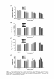
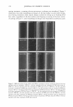
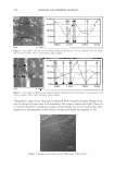
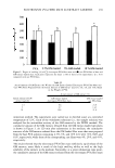
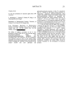
CHANGES IN HAIR DURING PERMANENT WAVING 205 For physicomorphological examinations, virgin hair samples (approximately 20 cm long) were also obtained from the same woman. Hair sample shafts were cut 15 cm from the scalp to provide hairs with minimal damage. The hair specimens with no chemical treatment were used and an expert with more than ten years of experience performed this experiment. Twenty pieces of the hair shafts of 80-89 µm diameter were grouped into one bundle (Figure 1). Each treatment consisted of three bundles. The prepared bundles were first rinsed with a neutral shampoo solution (pH 7 .8), washed with distilled water, dried, and stored in desiccators. The temperature and relative humidity of the desiccators were adjusted to 20°C and 65% for further use. PROTEIN EXTRACTION FROM HUMAN HEAD HAIRS After the collected hair samples were prepared after three washings using distilled water, the protein was extracted in a buffer solution containing 25 mM Tris-HCl (pH 8.5), 2.6 M thiourea, 5 M urea, and 5% 2-mercaptoethanol. The total protein was extracted by placing the hair materials into a tube or container containing the buffer solutions. After each sample group had been washed, 20 mg of each of the hair materials was incubated in the buffer solution (5 ml per 20 mg materials) at 50°C for 24 hr. After completion of their incubations, the mixtures were filtered through three layers of nylon mesh (200-mesh size) and a flow-through was used for the protein measurement. The detailed extraction method is described elsewhere (12). PROTEIN MEASUREMENT The amount of protein was measured with the protein-dye binding method of Bradford (13) using a commercial protein assay kit (Bio-Rad, Hercules, CA). For this analysis, 10 µl of the flow-through filtrate was transferred to a cuvette containing 200 µl of dye solution and 790 µl of distilled water to make 1000 µl in total. The amount of the total protein secreted into the buffer solutions was then quantitated at an optical density of 595 nm with a spectrophotometer (HP Agilent 8453, Palo Alto, CA). Each treatment was the mean value±standard deviation calculated in triplicate. SDS-PAGE EXPERIMENT For analysis of sodium dodecyl sulfate-polyacrylamide gel electrophoresis (SDS-PAGE), Figure 1. Features of human head hair samples prepared by different treatments. Twelve bundles including the control were used. Each wisp consists of 20 pieces of the hair shafts with 80-90 µm diameter.
Purchased for the exclusive use of nofirst nolast (unknown)
From: SCC Media Library & Resource Center (library.scconline.org)