]. Cosmet. Sci., 59, 225-232 (May/June 2008) AFM capabilities in characterization of particles and surfaces: From angstroms to microns N. STAROSTINA, M. BRODSKY, S. PRIKHODKO, C. M. HOO, M. L. MECARTNEY, and P. WEST, Pacific Nanotechnology Inc., 17981 Sky Park Circle, Irvine, CA 92614 (N.S, P. W.), University of California Los Angeles, UCLA Center for East-West Medicine, Los Angeles, CA 90095-1736 (M.B.), University of California Los Angeles, Department of Materials Science and Engineering, Los Angeles, CA 90095-1595 (S.P.), and University of California Irvine, Chemical Engineering and Material Science, Irvine, CA 92627-2575 (C.M.H., M.L.M.). Accepted for publication January 8, 2008. Presented at the Annual Scientific Meeting and Technology Showcase of the Society of Cosmetic Chemists, New York, December 7-8, 2006. Synopsis Scanning probe microscopy (SPM), invented 25 years ago, is now routinely employed as a surface charac terization technique. Atomic force microscopy (AFM) is the most widely used form of SPM, since AFM can be used in ambient conditions with minimal sample preparation. Examples of applications relevant to cosmetics include, but are not limited to, hair and skin roughness measurements and powder particle and nano-emulsion characterization. AFM is well suited for individual particle characterization, especially for measurements of volume, height, size, shape, aspect ratio, and particle surface morphology. Statistical distributions for a large set of particles can be generated through single-particle analysis techniques (i.e., ensemble-like information). AFM is better capable of resolving complex particle-size distributions than dynamic light-scattering (DLS). Single-particle analysis techniques with AFM can be more cost- and time-effective than analyses using scanning electron microscopy (SEM). However, AFM offers resolution that is comparable to or greater than SEM or transmission electron microscopy (TEM) and routinely allows direct measurements of the particle height and volume and produces images easily displayed in a quantified 3D format. INTRODUCTION AFM has been successfully employed for surface topography characterization over the last two decades. IBM researchers developed scanning tunneling microscopy (STM), which is considered an antecedent to AFM, in 1982 (1,2). AFM was invented in 1986 Address all correspondence to N. Starostina at Emerson Process Management, Rosemount Analytical Inc., Liquids, 2400 Barranca Parkway, Irvine, CA 92606. 225

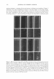
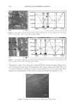
226 JOURNAL OF COSMETIC SCIENCE in order to overcome the limitations of STM and broadened the base of applications for SPM. Historically, the field of material science was the first to embrace the technology for nanoscale measurements (1-4). The range of the applications in particulate matter reaches from nanoparticle morphology visualization to quantitative characterization of a single particle to ensemble-like measurements of complex distributions (5). The AFM technique percolated into adjacent fields of colloidal chemistry, biology, biochemistry, and biomedicine. Currently it is being utilized in dentistry and the pharmaceutical industries. Phenomena regarding particle conformation, particle particle interaction, agglomeration, and adhesion have recently attracted the attention of many researchers in these fields (6-8). AFM structural investigations of viruses in solution have brought to light many answers that had been out of reach by other techniques (9). AFM studies of tooth enamel at the nanoscale range has been one focus of interest for dentistry (10,11). The goal of this paper is to show how AFM capabilities that have been traditionally used in material science can now be employed in the cosmetics industry to advance product development. A more comprehensive review of AFM can be found in the literature (3,4,12). In brief, AFM application advantages and capabilities can be summarized as follows: (a) Particles and surfaces can be imaged and characterized quantitatively in ambient conditions, in intervening media including liquids, or in an ultra-high vacuum (UHV). (6) Electric conductivity of the samples is not required and sample preparation is minimal (13). (c) Physical properties of materials, such as magnetic, electrical, thermal, and mechanical parameters, can also be measured. EXPERIMENT AL REPLICA SPECIMEN A negative replica of the skin surface was made using silicon rubber material mixed with a catalyst (Xantropen L Blue with Cuttersil). Replicas overlying the glabella were taken at baseline and repeated after eight weekly AcuFacial treatments at the UCLA Center for East-West Medicine. AcuFacial treatments included acupuncture, Chinese-style facial massage, and patient education on self-acupressure. Lifestyle aspects such as stress man agement, exercise, sleep, and diet were also addressed. Information on the different levels of effectiveness of the various topical prescription and over-the-counter anti-aging prod ucts on the market enabled patients to make informed decisions about the use of these products. For both the baseline and post-treatment replicas, the subject was placed in a supine position. Eyes were closed, and contractions of facial muscles were avoided. A replica ring locator (Replica™ Standard Replica Locator) was placed overlying the glabella, using the root of the nose and eyebrow ridges as landmarks. On a clean tray, the silicon rubber material was mixed together with the catalyst for 15-20 seconds in a ratio of 1 drop of catalyst per 1 ml of rubber material. The 1-2 ml of mixture paste was smeared over the replica to a final thickness of 2-3 mm. After one minute of polymerization, the impression was removed together with the replica ring. Orientation of the replica ring
Purchased for the exclusive use of nofirst nolast (unknown)
From: SCC Media Library & Resource Center (library.scconline.org)