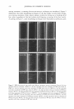
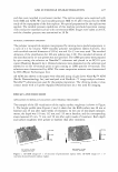
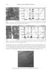
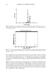
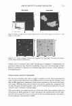
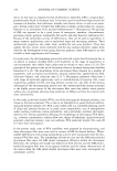
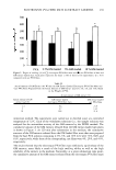
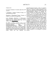
AFM IN PARTICLE CHARACTERIZATION 231 Particle Anal,- Particle Analylla Figure 8. Height distribution for foundation powder particles: 40 x 40 µm image is on the left 3 x 3 µm image is on the right. .. • • Figure 9. 20 x 20 µm topography (left-hand side) and phase mode (right-hand side) images of foundation powder. Images obtained simultaneously. Powder particle evaluation data show morphological parameters complemented by qualitative elasticity data. Particle morphology along with the knowledge of their physical parameters is important for manufacturing process control, surface chemistry, and agglomeration. CONCLUSIONS AND FUTURE WORK Pre- and post-treatment pilot data on surface roughness and line depth measurements from a single patient demonstrate that AFM may be utilized to measure changes in the skin to offer quantitative assessment of anti-aging treatments at the micron and nano scale ranges. AFM data from a future larger trial could be validated by comparison with existing descriptive and photographic scales, as well as profilometry techniques, that are currently used to evaluate cutaneous aging. The reported data on bi-modal particle size distribution of the aqua-polymer suspension demonstrate an AFM capability to resolve adequately the nominal peaks for the nano particles constituting the mixture. Individual as well as ensemble particle data analysis can be performed with the help of AFM.

232 JOURNAL OF COSMETIC SCIENCE Applications for AFM in the cosmetic industry include rapid and highly accurate par ticle size characterization and surface roughness measurements. The major advantages of using AFM in the cosmetic industry are ease of use, direct quantitative data along with understandable images, and minimal sample preparation. REFERENCES (1) G. Binning, C. F. Quate, Ch. Gerber, and E. Weibel, Surface studies by scanning tunneling micros copy, Phys. Rev. Lett. 49, 57-61 (1982). (2) G. Binning, C. F. Quate, and Ch. Gerber, Atomic force microscopy, Phys. Rev. Lett., 56(9), 930-933 (1986). (3) D. Bonnell, Scanning Probe Microscopy and Spectroscopy (Wiley-VCH, New York, 2001). (4) P. West and N. Starostina, Atomic force microscopy, Adv. Mater. Proc., 35-37 (February 2004). (5) N. Starostina and P. West, Tip dilation and AFM capabilities in the nanoparticle characterization,]. Mater., 12-16 Qanuary 2007). (6) T. Dukette and M. Mackay, Conformation of intramolecularly cross-linked polymer nanoparticles on solid substrates, Nano Lett., 5(9), 1704-1709 (2005). (7) K. Liu, Deformational behavior of soft particles: A Review,]. Phys. D: Appl. Phys., 39, Rl89-R199 (2006). (8) J.C. Hooton, C. S. German, S. Allen, M. C. Davies, C. J. Roberts, S. J. B. Tendler, and P. M. Wil liams, An atomic microscopy study of the effect of nanoscale contact geometry and surface chemistry on the adhesion of pharmaceutical particles, Pharmaceut. Res., 21, 953-961 (2004). (9) Yu. Kuznetsov, J. Gurnon, J. Van Etten, and A. McPherson, Atomic force microscopy investigation of a chlorella virus, PBCV-1,j. Struct. Biol., 149, 256-263 (2005). (10) C. Robinson, K. Yamamoto, S. D. Connell, J. Kirkham, H. Nakagaki, and A. D. Smith, The effects of fluoride on the nanostructure and surface pK of enamel crystals: An atomic force microscopy study of human and rat enamel, Eur.]. Oral Sci., 114(Supl.1), 99-104 (2006). (11) F. Watari, In situ quantitative analysis of etching process of human teeth by atomic force microscopy, ]. Electron Microsc., 54(3), 299-308 (2005). (12) N. Burnham, R. Colton, and H. Pollock, Interpretation issues in force microscopy,]. Vac. Sci. Technol., A 9(4) Quly/August 1991). (13) N. Starostina and P. West, AFM capabilities in characterization of particulate matter: From angstroms to microns, Proceedings of the 33rd Annual International Waterborne, High-Solids, and Powder Coating Symposium, 307-320 (February 2006). (14) P. Goodhew, J. Humpreys, and R. Beanland, Electron Microscopy and Analysis (Taylor & Francis, 2001).
Purchased for the exclusive use of nofirst nolast (unknown)
From: SCC Media Library & Resource Center (library.scconline.org)