JOURNAL OF COSMETIC SCIENCE 386 it has long been recognized that skin and other epithelia can produce a range of antimicrobial peptides that play an important part in eliminating potential cutaneous pathogens (6,7). Antimicrobial peptides are induced in keratinocytes of the skin via the binding of bacte- rial lipopolysaccharides, beta glucans, some mannose-containing carbohydrates, peptido- glycans or bacterial DNA to “pattern recognition” receptors, or the toll-like receptors. Heat-killed bacteria or fungi alone can provoke the transcription of antimicrobial pep- tides. Lactobacillus and other types of lactic acid bacteria have been reported to produce specifi c antimicrobial peptides known as bacterocins (8) that possess broad-spectrum an- timicrobial activity against gram-negative and gram-positive bacteria (9). Oral administration of probiotics has been shown to be effective in improving skin barrier functions (10). Impaired skin barrier function is invariably caused by decreased amounts of ceramides that may be responsible for comedone formation, since barrier dysfunction is accompanied by hyperkeratosis of the follicular epithelium (11). Since microbial agents, impaired barrier, and infl ammation play an important role in acne, we considered observing the effect of a probiotic on acne. Acne vulgaris is a com- plex, chronic, and common skin disorder of pilosebaceous units (12). The major patho- genic factors involved are ductal hyperkeratinization, obstruction of sebaceous follicles resulting from abnormal keratinization of the infundibular epithelium, stimulation of sebaceous gland secretion by androgens, and microbial colonization of pilosebaceous units by Propionibacterium acnes. Both viable and non-viable P. acnes have been shown to induce an immunostimulatory effect that activates an infl ammatory response (13). The infl ammatory stage of acne vulgaris is usually of greatest concern to the patient. A num- ber of morphologically different infl ammatory lesions may form that can be painful and unsightly and can lead to scarring. Infl ammatory acne and acne scarring can have signifi - cant psychological effects on the patient, including depression, anxiety, and poor self- image. The onset of non-infl ammatory lesions is understood as the consequence of follicular keratinocytes failing to differentiate, thus producing hypergranulosis, resulting in the formation of microcomedones (12). Acne is commonly treated with antibiotics, bactericidals, retinoids, and so on, however, most of such treatments come with side effects. In this study, we addressed alleviation of three aspects of acne, namely skin microfl ora, barrier strength, and infl ammation using a probiotic: lactobacillus extract. MATERIALS AND METHODS The test material was the probiotic lactobacillus extract (14). The Lactobacillus ferment was prepared in MRS broth (Neogen Corporation, Lansing, MI) inside a 1000-L fermen- ter sterilized for 20 min at 121°C to insure sterility. The vessel was cooled to 37°C and nitrogen was pumped into the vessel until the total dissolved oxygen was down to zero in order to ensure anaerobic conditions for the growth of Lactobacillus plantarum. The vessel was inoculated with 10 L (or 1% of total volume) of L. plantarum prepared in the same MRS media broth and same conditions as the fi nal 1000-L vessel. The organism was al- lowed to grow in the vessel for 18–24 h after which the ferment was passed through a heat exchanger to lyse most of the cells. The ferment was then fi ltered fi rst through a 0.45-μm fi lter, and fi nally through a 0.22-μm fi lter to produce the fi nal broth (14).

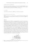
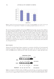
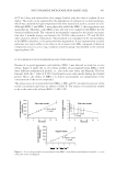
PHYSIOLOGICAL EFFECT OF A PROBIOTIC ON SKIN 387 Oil-in-water (o/w) formulations were prepared at 1% and 5% concentrations of the pro- biotic for the studies. Triclosan (BASF, NC), a known antibacterial, at 0.1% concentra- tion prepared in a similar formulation base, was used as a control for some parts of the study. In addition, a similar formulation base with 1% salicylic acid (Rhodia Inc., NJ) was used as an internal control for the acne study presented in this paper. All clinical studies were conducted following good clinical practice standard (ICH Topic E 6-R1 July 2002 CPMP/ICH/135/95). The subjects recruited in these studies were in normal health with no evidence of acute or chronic disease other than acne. Written in- formed consent was obtained from each volunteer before entering into the study. The subjects were not on any antibiotic, antihistamines, retinoid, anti-infl ammatories or steroid therapy, benzoyl peroxide, and/or salicylic acid treatment for at least 2 weeks prior to commencement of this study. The subjects were not under the care of a der- matologist and were not on any acne treatment for at least 1 month before the study started. Pregnant or lactating females were excluded, also subjects exhibiting current sunburn, rashes, scratches, burn marks, etc., which might interfere with the evaluation of test results. SKIN SENSITIVITY The anti-infl ammatory properties of the test materials were tested by observing the re- duction of onset and intensity of erythema induced by an irritant, Balsam of Peru. The test site was on the volar forearms of subjects with a history of sensitivity to Balsam of Peru (15). “Balsam of Peru” (8% w/w in petrolatum), an irritant that contains approxi- mately 0.9% cinnamic aldehyde (16,17), was applied at a dose of approximately 4 mg/cm2. Erythema was measured with a Minolta Chromameter (Konica Minolta, Ramses, NJ). Part I: Reduction of onset of skin redness. Ten subjects with a history of skin sensitivity to Balsam of Peru were chosen for the study. The test compounds were applied on the volar forearms of the subjects. The material was allowed to absorb for 30 min and then Balsam of Peru, the irritant, was applied on the test sites. When erythema appeared, even on one site, the arms were washed with wet towels and skin redness was measured with the Chromameter. Red (a∗ values) subtracted by the baseline skin redness determined an “increase in redness (Δa∗) due to irritation.” A comparison of Δa∗ with the positive and negative controls exhibited the potential of the test materials for reducing the onset of skin irritation. The positive control was a∗ values of skin treated with Balsam of Peru alone. Part II: Reduction of intensity of skin redness. A total of ten subjects participated in the study. Using a pen, 1.5 in.2 areas were marked on each volar forearm of the subjects correspond- ing to the test materials and the positive and negative controls. Baseline color measure- ments were obtained from all the sites using a Minolta Chromameter. Balsam of Peru (8% pet) was applied on all the sites at the rate of approximately 4 mg/cm2 in a 1.5-cm diameter circle. When redness appeared approximately evenly on all the sites, the irritant was wiped off with a wet towel and then washed with warm water. The degree of redness was measured with the Chromameter on all the sites as the baseline redness. The test materials were applied on their respective sites at the rate of 2 mg/cm2 and color measurements were obtained after 15 min, 30 min, 1 h, 1.5 h, and 2 h.
Purchased for the exclusive use of nofirst nolast (unknown)
From: SCC Media Library & Resource Center (library.scconline.org)