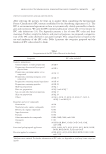
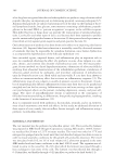
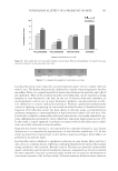
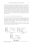
JOURNAL OF COSMETIC SCIENCE 388 Increase in color was determined by subtracting the baseline skin color values of all the sites and color values were plotted against time. Area under the curve was obtained for all the sites. SKIN BACTERIAL COUNT The test materials used in this section were an o/w base cream containing lactobacillus extract (L. plantarum) at 1% and a similar formulation with Triclosan (an antibacterial) at 0.1% as a control. A total of 29 females between the age of 25 and 55 participated in the study. The panel was divided into three groups of 9–10 each. The subjects were provided with the test material to use twice a day for 2 months on the full face. They were instructed not to use any other moisturizers or treatment products however, they could continue to use the cleansers and makeup that they normally use as long as they did not change products during the course of the study. On the day of the study, the subjects reported to the lab with a clean face and forearms, with no creams, lotions, makeup, etc. Skin microfl ora measurements described below were obtained at baseline, 1 month, and 2 months. Skin microfl ora. The subjects (n = 29) reported to the laboratory and washed their face with a (non-antibacterial) mild liquid soap. Normally, after washing with soap and water, the bacterial count of skin drops to almost zero with gradual increase in microfl ora over time. The normal microfl ora was allowed to appear on the skin for the next 3 h. During this pe- riod, the subjects were advised to keep their hair away from the forehead and refrain from touching the face or wash it or apply anything on it. At the end of this 3-h time point, saline washings were obtained from the forehead of the face for microbiological analysis. Dulbeccos phosphate-buffered saline (PBS) washings of the forehead area were obtained using a sterile glass cylinder and a sterile rubber policeman. One milliliter of the saline was poured in the cup and then the skin was scrubbed with rubber policeman (10 strokes) and washed and then the saline was aspirated and collected in 9 ml of PBS. The samples were analyzed for aerobic and anaerobic bacterial count. The data were averaged to deter- mine the total microfl ora. The samples were analyzed for microfl ora as per the U.S. Pharmacopeia Chapter 61 (18) where 1:10 dilutions of the samples were prepared in PBS and plated on tryptic soy agar. After 48 h of incubation at 37°C, the plates were examined and the recovered organisms were quantifi ed. All recovered organisms were then streaked for single colony isolation, gram stained, and examined under the microscope. The organisms were identifi ed using the Becton–Dickinson BBL Crystal™ Identifi cation System. BARRIER FUNCTIONS The test site was the jawline of the face of the subjects. Basal skin barrier was determined by measuring trans epidermal water loss (TEWL), using a Servomed Evaporimeter (Ser- voMed AB, Stockholm, Sweden). To determine barrier integrity, a tape (Tuck tape) was used to cover the test area and after a fi rm stroke in both directions the tape was peeled off (19). A total of three strippings were obtained. TEWL was recorded again. Strippings followed

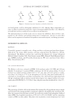
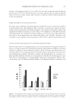
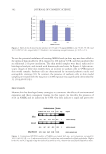
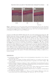
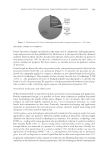
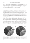
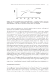
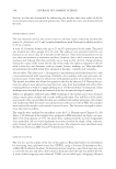
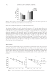
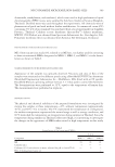
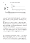
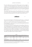
PHYSIOLOGICAL EFFECT OF A PROBIOTIC ON SKIN 389 by TEWL measurements were continued in groups of three. Damage to the barrier was described as TEWL of 18 g/m2/h. The number of strippings to disrupt skin barrier was calculated using a linear equation. Barrier repair was observed by measuring TEWL at the stripped skin site, 3 h and 24 h after stripping. For each subject, a linear graph was plotted for TEWL versus time (hours) and using a linear equation, the number of hours to reduce disrupted barrier TEWL to 50% was calculated for every time point (15, 19–21). The subjects used an o/w base cream containing lactobacillus extract (L. plantarum) at 1% for 2 months and the same measurements were observed after 1 and 2 months of use. ACNE LESION REDUCTION Oil-in-water base formulations containing lactobacillus extract at 1% and 5% were tested against a formulation containing 1% salicylic acid as a control. Ten volunteers between the age of 18 and 50 were recruited from a local population. The subjects exhibited acne with several similar acne lesions on the upper back. Two infl amed acne lesions were selected for each treatment and for the untreated. Each lesion was marked, photographed, and graded. A skin surface microscope (Scopeman, Moritex U.S.A., Inc., San Jose, CA) was used to visualize, size, and grade the lesion by two MDs at the testing labora- tory. The lesions were treated with the respective treatment once a day for 4 days. Acne le- sion size and erythema were plotted against time and area under the curve was calculated (22), and statistical signifi cance was calculated using students t-test. RESULTS SKIN SENSITIVITY: EFFECT OF LACTOBACILLUS EXTRACT ON THE REDUCTION OF THE ONSET OF SKIN IRRITATION As observed in Figure 1, there was 57% and 40% reduction in the onset of erythema with 5% and 1% lactobacillus extract, respectively, showing a dose response. Figure 1. Effect of lactobacillus extract (in o/w emulsion) on the reduction of the onset of skin irritation.
Purchased for the exclusive use of nofirst nolast (unknown)
From: SCC Media Library & Resource Center (library.scconline.org)