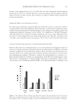
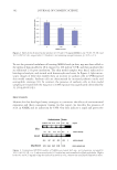
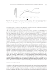
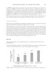
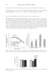
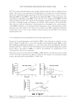
JOURNAL OF COSMETIC SCIENCE 400 The chemical stability at 60°C was not anticipated since in the guideline for accelerated stability test of new drug substances and products suggested by International Conference on Harmonization (17), the studied temperature is assigned at 40°C. Physical changes of the samples such as phase separation and clarity were optically observed. Only MBG-1 was selected to investigate for the chemical stability study by determining the amount of non-degraded active compound using HPLC. The 100 μl of the formulation was dis- solved with IPA and adjusted the volume in a 10-ml volumetric fl ask. The 100 μl of this solution was diluted with 80:20 IPA/methanol in a 10-ml volumetric fl ask. Afterward, the obtained solution was further diluted with the same solvent in a 10-ml volumetric fl ask and then fi ltered through a 0.45-μm polyamide membrane fi lter before HPLC analysis. IN VITRO RELEASE STUDY The release of nicotinamide from MBG-1, ME, and CC was investigated using modifi ed Franz diffusion cells (Hanson Model 57-6 M, Hanson Research Corporation, Los Angeles, CA) and a cellulose acetate membrane with an effective diffusion area of 1.77 cm2. The membrane was cut into 3 × 3 cm pieces, and the obtained pieces were boiled in distilled water to remove the wax. The cleaned membrane pieces were soaked in distilled water, stored in a cool place, and used within a week. The hydrated membrane pieces were mounted between the donor and receptor compartments of the diffusion cells. The receptor compart- ment was fi lled with 11 ml of degassed IPB. The diffusion cells were connected to a circu- lating water bath thermostated at 37°C, giving the membrane surface temperature of 32°C that was equal to human skin temperature. Each diffusion cell was continuously stirred at a speed of 200 rpm using a magnetic bar. After the membrane was equilibrated for 30 min, 1 g of each formulation was placed onto the membrane in the donor compartment. The donor compartment and the sampling arm were covered with Parafi lm to prevent water evaporation. At defi ned time intervals (0.5, 1, 2, 4, 8, 10, 12, and 25 h), 0.5 ml of samples were taken from the receptor compartment and immediately replaced with an equal volume of fresh receptor medium. The withdrawn samples were analyzed for nicotinamide concen- tration by HPLC. For each formulation, the experiment was performed in triplicate. The cumulative release (Q) of nicotinamide was calculated from equation (1). ¦V 1 0 t r t s i i Q V C C (1) where Ct is the concentration of active compound in the receptor fl uid at each sampling time t, Ci is the concentration of active compound of the ith sample, and Vr and Vs are the volumes of the receptor fl uid and the sample, respectively. The release rate was calculated as percent of the initial concentration per hour by linear regression interpolation of the experimental data. Three possible mathematical equations were employed to fi t with the release profi les, i.e., zero order, fi rst order, and Higuchi square root of time equations as shown in equations (2), (3), and (4), respectively.

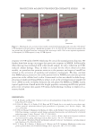
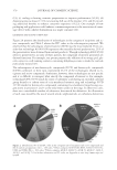
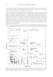
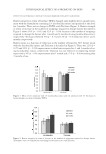
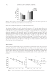
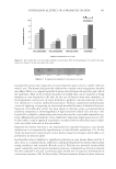
NICOTINAMIDE MICROEMULSION-BASED GELS 401 0 0 Q Q k t (2) 0 ln lnQ f Q k (3) 1/2 H Q k t (4) where Q is the cumulative amounts of active compound released in time t Q0 is initial amounts of active compound in the preformed preparations and k0, kf, and kH are release rate constants of zero order, fi rst order, and Higuchi model, respectively (18). HPLC ASSAY The concentrations of nicotinamide remained in MBG-1 after stability study and released from the investigated samples into the receptor fl uid were quantitatively analyzed by HPLC as described by Xu and Trissel (19) with some modifi cations. The HPLC system (Agilent 1100, Santa Clara, CA) connected with an HPLC column, 5-μm particle size, 150 × 4.6 nm (Chrompack Inertsil ODS, Middelburg, The Netherlands). A mixture of 0.1% triethylamine in 0.067 M monobasic potassium phosphate buffer (pH 6.7) and acetonitrile (100:4 v/v) was used as the mobile phase. The injection volume was 20 μl. The samples were detected at 260 nm and integrated with the RF 10A (version 1.1) LC software program. The calibration curve was constructed by running standard solutions of nicotinamide in extraction solvent and in IPB for every series of samples. Validation of the method was performed to ensure that chromatogram of the standard solution of nico- tinamide could be separated from chromatogram of the blank MBG extract and that of receptor fl uid incubated with cellulose membrane. The calibration curve between 2.5 and 40 μg/ml of nicotinamide was in the linearity range (r2 0.999) and coeffi cients of varia- tion were less than 2%, both intraday and interday. RESULTS AND DISCUSSION CHARACTERISTICS OF NICOTINAMIDE MICROEMULSION-BASED GELS According to optical observation, the rank order of clarity of the prepared MBGs was MBG-1 MBG-3 MBG-2. It was found that using colloidal silica provided a transpar- ent gel. The result can be explained that during the gel formation, H+ ions of water at- tached to some of the small particles of colloidal silica, resulting in the hydrophilic surface and capability of hydrogen bonding. This structure formed into a three-dimensional net- work through the branched interaction of hydrogen bonding of hydroxyl groups on silica surface (20,21). Although the water in the system was low, the water cores of ME might serve as compartmentalized media for this reaction due to their dynamic characteristics (22). Colloidal silica was previously reported that it converts sodium ascorbyl phosphate
Purchased for the exclusive use of nofirst nolast (unknown)
From: SCC Media Library & Resource Center (library.scconline.org)