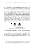
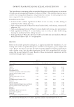
BOTANICALLY DERIVED SKIN SURFACE LIPID MIMETIC 67 REFERENCES (1) N. Nicolaides, “Human skin surface lipids—origin, composition and possible function” in Advances in Biology of Skin, W. Montagna, R. A. Ellis, and A. F. Silver. Eds. (Pergamon Press, Oxford, England, 1963), pp. 167–186. (2) R. S. Greene, D. T. Downing, P. E. Pochi, and J. S. Strauss, Anatomical variation in the amount and composition of human skin surface lipid, J. Invest. Dermatol., 54, 240–247 (1969). (3) P. M. Elias, Epidermal lipids, barrier function, and desquamation, J. Invest. Dermatol., 80 (Suppl. 1), 44s–49s (1983). (4) A. M. Porter, Why do we have sebaceous glands?, J. R. Soc. Med., 94, 236 (2001). (5) A. Kydonieus and J. J. Wille, Palmitoleic acid isomer (C16: 1Δ6) in human skin sebum is effective against gram-positive bacteria, Skin Pharmacol. Appl. Skin Physiol., 16, 176–187 (2003). (6) U. Jacobi, J. Gautier, W. Sterry, and J. Lademann, Gender-related differences in the physiology of the stratum corneum, Dermatology, 211, 312–317 (2005). (7) A. M. Kligman and D. L. Miller, Sebutape: A device for visualizing and measuring human sebaceous secretion, J. Soc. Cosmet. Chem., 37, 369–374 (1986). (8) J. S. Strauss and P. E. Pochi, The quantitative gravimetric determination of sebum production, J. Invest. Dermatol., 36, 293–298 (1961). (9) A. Anderson and J. Fulton, Sebum: analysis by infrared spectroscopy, J. Invest. Dermatol., 60, 115–120 (1973). (10) R. Michael-Jubeli, A. Tfayli, J. Bleton, and A. Baillet-Guffroy, Chemometric approach for investigating the skin surface lipids (SSLs) composition: Infl uence of geographical localization, Eur. J. Dermatol., 21 (Suppl. 2), 63–71 (2011). (11) L. C. Robosky, K. Wad, D. Woolson, J. D. Baker, M. L. Manning, D. A. Gage, and M. D. Reily, Quan- titative evaluation of sebum lipid components with nuclear magnetic resonance, J. Lipid Res., 49, 686– 692 (2008). (12) E. Camera, M. Ludovici, M. Galante, J. L. Sinagra, and M. Picardo, Comprehensive analysis of the major lipid classes in sebum by rapid resolution high-performance liquid chromatography and electrospray mass spectrometry, J. Lipid Res., 51 (11), 3377–3388 (2010). (13) A. Pappas, J. Fantasia, and T. Chen, Age and ethnic variations in sebaceous lipids, Dermato-Endocrinology, 5, 319–324 (2013). (14) S. S. Shetage, M. J. Traynor, M. B. Brown, M. Raji, D. Graham-Kalio, and R. P. Chilcott, Effect of ethnicity, gender and age on the amount and composition of residual skin surface components derived from sebum, sweat and epidermal lipids, Skin Res. Technol., 20, 97–107 (2014). (15) J. A. Cotterill, W. J. Cunliffe, and B. Williamson, Age and sex variation in skin surface lipid composi- tion and sebum excretion rate, Br. J. Dermatol., 87, 333–340 (1972). (16) I. B. Ro and T. L. Dawson, The role of sebaceous gland activity and scalp microfl oral metabolism in the etiology of seborrheic dermatitis and dandruff, J. Invest. Dermatol. Symp. Proc., 10, 194–197 (2005). (17) I. Buraczewska, B. Berne, M. Lindberg, H. Törmä, and M. Lodén, Changes in skin barrier function following long-term treatment with moisturizers, a randomized controlled trial, Br. J. Dermatol., 156, 492–498 (2007). (18) P. Todorova, P. Grant-Ross, S. Tamburic, and R. Kurimo, Biomimetic vs. traditional skin moisturiza- tion: An in vivo comparison, Cosmetics Toiletries., 130, 30–40 (2015). (19) R. Michael-Jubeli, J. Bleton, A. Baillet-Guffroy, High-temperature gas chromatography-mass spec- trometry for skin surface lipids profi ling, J. Lipid Res., 52, 143–151 (2011). (20) P. Ramasastry, D. T. Downing, P. E. Pochi, and J. S. Strauss, Chemical composition of human skin surface lipids from birth to puberty, J. Invest. Dermatol., 54, 139–144 (1970). (21) A. V. Rawlings, Ethnic skin types: Are there differences in skin structure and function, Int. J. Cosmet. Sci., 28, 79–93 (2006). (22) J. M. Ntambi, “Stearoyl-CoA desaturases are regulators of lipid metabolism in skin” in Lipids and Skin Health, A. Pappas Ed. (Springer Science: New York, NY, 2015), pp. 239–248. (23) M. M. Downie and T. Kealey, Lipogenesis in the human sebaceous gland: Glycogen and glycerophos- phate are substrates for the synthesis of sebum lipids, J. Invest. Dermatol., 111 (2), 199–205 (1998). (24) B. Middleton, I. Birdi, M. Heffron M, and J. R. Marsden, The substrate determines the rate and pattern of neutral lipid synthesized by isolated human sebaceous glands, FEBS Lett., 231 (1), 59-61 (1988).

J. Cosmet. Sci., 68, 68–73 ( January/February 2017) 68 Address the epidemic of p-phenylenediamine sensitization and aid marginal farmers in a changing climate CATHERINE CARTWRIGHT-JONES, TapDancing Lizard LLC. THE EFFECT ON THE HAIR DYE INDUSTRY OF THE EPIDEMIC OF P-PHENLYLENEDIAMINE SENSITIZATION There is a global epidemic of sensitization to p-phenylenediamine (PPD) (1). The popularity of using black hair dye to create “black henna” temporary tattoos as vacation souvenirs for Westerners as well as for Muslim and Hindu weddings and social occasions has exposed millions of people to highly sensitizing doses of PPD. Of these people, 50% become allergic to oxidative hair dye and 20% become severely allergic (2). The most commonly used materials for black henna tattoos are 15–40% PPD black hair dye powders (3) as well as chunks of 90% + pure industrial PPD (4). The ornate black henna patterns cover large skin surface areas and the black henna paste is left in place for half an hour or more intro- ducing a dangerously high dose of PPD to the body. Substitution of PPD for safe traditional henna body art began in east Africa in the 1970s when Bigen and Peacock home hair dye kits were marketed in the region. This practice spread and became fashionable in Saudi Arabia, Egypt, Pakistan, and India by the early 1980s (5). Women used high PPD content black oxidative chemical hair dye powders for body art instead of henna because PPD produces black stains on skin quickly the fi nely powdered chemical dye can be manipulated into more complex patterns than roughly sifted local henna. PPD was applied to skin of brides and their wedding guests, and was used for social celebrations. Many were sensitized by the fi rst time their skin was painted with PPD if they were not sensitized in the fi rst application, subsequent celebrations with black henna for Eids, Karva Chauth, and Diwali sensitized them. Five or fewer applica- tions of 10% PPD in a patch test will sensitize 100% of subjects (6) black henna contains 15–60% PPD. Some were unaware that PPD sensitization would affect their health in the future others regarded the blisters that arose after application to be of little concern, regarded as “suffering for beauty.” They felt black patterns were more beautiful on darker skin as well as more convenient. As these women mature, they may decide to dye their hair with oxidative hair dye. For these women, the hypersensitivity reaction can be severe anaphylaxis may be fatal (7). Forty women have died in recent years in Libya from PPD sensitization (8), and offi cials have called for a ban on oxidative hair dye. There has been Address all correspondence to Catherine Cartwright-Jones at reverndbunny@earthlink.net.
Purchased for the exclusive use of nofirst nolast (unknown)
From: SCC Media Library & Resource Center (library.scconline.org)