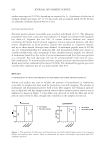
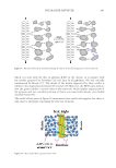
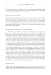
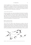
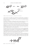
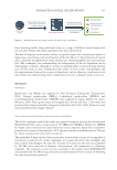
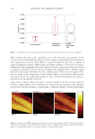
698 JOURNAL OF COSMETIC SCIENCE The different chemical mechanisms causing these changes are complex and difficult to measure directly in hair. There is strong evidence that two key amino acids in hair that absorb UV-B radiation are tryptophan and tyrosine. These amino acids are photoionized and produce aromatic free radicals. Tryptophan oxidation produces yellow-colored kynurenines (5), which can be observed as a photo-yellowing effect in light-colored hair. Singlet oxygen and a superoxide radical anion are formed by the photosensitization of both aromatic amino acid residues and melanin pigments (6). Cell membrane complex unsaturated lipids such as the unsaturated fatty acids react with singlet oxygen to form hydroperoxides that modify the cell membrane complex and provide hydroxyl and alkoxyl radicals for additional reactions. These reactions have been studied and reported in both hair and wool (2,7). Reactive oxygen species (ROSs) that are formed continue to propagate damage throughout the hair. It has also been shown that redox metals such as copper can accelerate these radical reactions and create additional protein damage (8). An approach to reducing UV damage is to terminate the reactions by quenching free radicals or ROSs. Primary antioxidants, such as polyphenols, act in this manner. There are three main mechanisms by which antioxidants can scavenge ROSs: hydrogen-atom transfer, single-electron transfer, and metal chelation. Polyphenols can act as antioxidants by these mechanisms and are important because they are commonly found in botanical extracts including tea (Camellia sinensis) (9). Several publications have studied the use of tea extract for hair and skin benefits (10), including as protection against UV-induced skin damage and in the development of sunscreen products (11). Tea extracts have also been studied for their impact on hair growth (12) and sebum reduction (13). The objective of this study was to create a model system for the key components in hair involved in the initiation of radical pathways and gain a more detailed understanding of specific pathways involved, including the role of redox metals. From this work, the strategy of adding antioxidants was identified as an option for reducing oxidative damage. A selection of tea extracts that can prevent UV damage by intercepting the ROSs formed were screened and shown to reduce this oxidative damage. MATERIAL AND METHODS POLY(ETHYLENE GLYCOL)–TYROSINE BLOCK COPOLYMER MICELLES Poly(ethylene glycol)–tyrosine (PEG–Tyr) polymers (prepared according to published literature procedures [14]) were kindly donated by Prof. A. Heise, Dublin City University, Dublin, Ireland (14). PEG 5000 –Tyr 15 (20 mg, 0.00259 mmol), PEG 2000 –Tyr 5 (10 mg, 0.00344 mmol), and PEG 5000 –Tyr 10 (20 mg, 0.00293 mmol) were separately added to water (1 mL). The mixtures were sonicated at room temperature for 60 min to ensure homogenous dissolution into colloidal systems. The colloidal systems were irradiated using a 100 W mercury arc lamp (Oriel 6281/ Ushio USH-102DH) with spectral irradiance ranging from deep UV through infrared wavelengths. To add iron(III) stearate and iron(III) acetylacetonate to the polymer, stock Fe(III) solutions were prepared. Specific volumes of the stock solution were transferred into sample vials to give the desired metal concentration when diluted by a factor of 100. The ethanol was then removed under a vacuum, and PEG 2000 –Tyr 5 polymer solutions (0.5–5 mg/mL, 0.074– 0.735 mM) were prepared in 0.1 M pH 5 acetate buffer and added to dried Fe residue. The

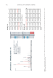
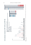
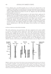
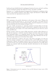
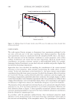
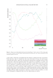
699 UV OXIDATION resulting solutions were shaken for 1 h, sonicated for 1 h, and then shaken for an additional 1 h to ensure partitioning of the hydrophobic metal salt into the colloidal system. FLUORESCENCE ANALYSIS OF COLLOIDAL SYSTEMS After each sample had been irradiated, 150 μL of ethanol was added to dissolve any aggregates. Then, 200 μL of the resulting solution was withdrawn, and 400 μL of each 0.1 M pH 5 acetate buffer and 0.4 M pH 8.5 borate buffer was added. This decreased the ethanol concentration sufficiently for micelle structures to form, as tested by dynamic light scattering. Fluorescence excitation and emission spectra were recorded, with an average of three scans taken for each sample. (Excitation parameters: emission wavelength, 303 nm excitation scan, 240–300 nm. Emission parameters: excitation wavelength, 276 nm emission scan, 270–500 nm.) Dityrosine fluorescence was also monitored. (Excitation parameters: emission wavelength, 405 nm excitation scan, 240–400 nm. Emission parameters: excitation wavelength, 320 nm emission scan, 325–550 nm.) The extent of tyrosine degradation was measured as a percentage decrease in the tyrosine emission maximum intensity at 310 nm relative to the dark control for each sample. Fluorescence measurements were performed on a Hitachi F-4500 fluorescence spectrophotometer using 1 × 1 cm quartz cells. Samples were treated after irradiation to remove metals before fluorescence measurements were made. Iron was removed from the samples by the precipitation as Fe(OH) 3 at high pH: To 140 μL samples was added 150 μL of dilute NaOH solution (194 mM). Samples were shaken and left for 10 min for precipitation to occur and were then filtered through 450 nm polytetrafluoroethylene filters. BUTYLATED HYDROXYTOLUENE (BHT) EXPERIMENTS For studies with butylated hydroxytoluene (BHT) antioxidant, a stock BHT solution in ethanol at a concentration of 1.5 mM was prepared. Specific volumes of the stock solution were transferred into sample vials to give the desired BHT concentration when diluted by a factor of 100. The ethanol was then removed under a vacuum, and PEG 2000 –Tyr 5 polymer solutions (0.5–5 mg/mL, 0.074–0.735 mM) were prepared in 0.1 M pH 5 acetate buffer and added to dried BHT residue. The resulting solutions were shaken for 1 h, sonicated for 1 h, and then shaken for an additional 1 h to ensure partitioning of the hydrophobic antioxidant into the colloidal system. HAIR Human hair for the testing was purchased from International Hair Importers & Products, Glendale, New York. Natural white hair, a hair type that is very low in melanin content, was used for UV biomarker work. HAIR TREATMENTS For biomarker experiments, natural white hair was treated with solutions containing 5% C sinensis extract. These solutions were applied to wet hair and left to dry without rinsing. The resulting samples were compared to baseline untreated hair and UV-exposed-only
Purchased for the exclusive use of nofirst nolast (unknown)
From: SCC Media Library & Resource Center (library.scconline.org)