49 Address all correspondence to Elisabeth Stevens, lisa.ajdukovich@outlook.com Potential Blue Light Effects on Aging Mechanisms Within Adult Human Skin, a Literature Review ELISABETH STEVENS James L. Winkle College of Pharmacy, University of Cincinnati, Cincinnati, Ohio, USA (E.S.) Accepted for publication: November 7, 2021. Synopsis Blue light is high energy visible light in the region of 400–500 nm in wavelength. There have been questions raised in recent years as to whether this can influence skin in manners like UV light, often centered on the heavy widespread use of mobile devices. Blue light impacts skin through mechanisms such as inflammatory cytokines, cellular viability, and melanogenesis. According to current research conducted on in vitro cells as well as in vivo skin, it seems that blue light can have an effect on skin that can manifest as extrinsic aging, but this may only occur at the shorter end of the blue light spectrum and at levels of exposure that are sourced from the sun. INTRODUCTION BLUE LIGHT Types of light are categorized by wavelength. Ultraviolet C (UVC) is between 200–280 nm, ultraviolet B (UVB) is 280–320 nm, and ultraviolet A (UVA) between 320–400 nm. Visible light is within the 400–700 nm range (1). Blue light wavelengths have been reported as anywhere from 400–500 nm. Blue light can penetrate even further into the dermis than UVA and UVB light. In the dermis, blue light can potentially be absorbed by hemoglobin, riboflavin and flavoproteins, and other porphyrin-containing enzymes as found in their absorption peaks, which are in the blue light region (2). In the epidermis, melanin can absorb blue light (3). Fluence is defined as the wattage delivered per unit area with which an object is irradiated. Sunlight delivers a fluence of total radiation of 136 mW/ cm2 (1). However, about 40% of sunlight is reflected toward its source. Artificial sources of light provide only a fraction of the exposure levels as compared to the sun as a source. Exposure to a computer screen 18 in away for 10 min can deliver irradiance of 0.6 mW/cm2 to skin surface (4). J. Cosmet. Sci., 73, 49–57 (January/February 2022)
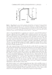
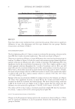
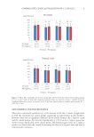
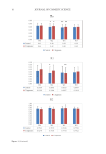
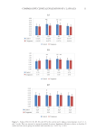
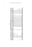
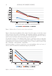
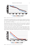
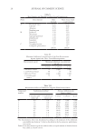
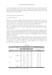
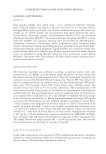
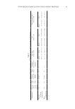
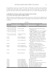
50 JOURNAL OF COSMETIC SCIENCE SKIN AGING There are two types of aging affecting the skin: (1) Intrinsic aging occurs mostly regardless of the levels of exposure of the skin to damaging elements at genetically determined rates and (2) extrinsic aging occurs because of external stressors (e.g., prolonged exposure to sunlight). Table I summarizes the main differences in how extrinsic and intrinsic aging manifest in the skin. BLUE LIGHTS IMPACT ON SKIN IN VITRO STUDIES Cell viability. Opländer studied different wavelengths’ effects on human dermal fibroblasts obtained from mammoplasty or abdominoplasty. Dermal fibroblast cell viability decreased at 410 and 420 nm at 60 and 90 J/cm2 (10,11). At 15 and 30 J/cm2, the reduction in viability was not statistically significant, and at 453 nm and higher, no significant reduction in viability was shown. Cells treated with hydrogen peroxide and blue light reduced cell numbers more than hydrogen peroxide alone, showing that blue light further reduces antioxidant activity of fibroblasts, since the reduction of cell numbers was increased when exposed to blue light. Wavelengths of 453 and 480 nm showed no change. Blue light at wavelengths 412–426 nm was toxic to endothelial cells and keratinocytes in a dose-dependent manner (as noted by Liebmann). Cells were irradiated at 0, 33, 66, and 100 J/cm2 every 24 h for 3 d, then 24 h after the last radiation, and the number of viable cells was measured using staining methods. The number of viable keratinocytes and endothelial cells varied inversely with the level of fluence delivered. Cytotoxicity was shown in both types of cells, although keratinocytes were more resistant. At 453 nm, there is no toxicity present at fluences up to 500 J/cm2, much higher than levels delivered by the sun (12). Melanogenesis. Regazzetti also studied blue light impact melanogenesis. Human foreskin obtained from children with skin types III and IV was used to culture melanocytes. Cells were irradiated with 415 nm blue light at 50 J/cm2. Using Western blot to test for proteins, there has been an increased calcium flux with blue light irradiated NHMs, and upregulation of pCAMKII. CAMKII activates CREB, then ERK, and p38 (13). This then phosphorylates melanogenesis-associated transcription factor (MITF), which then Table I A Summary of Aging Events for Extrinsic Versus Intrinsic Aging Types Skin layer Extrinsic aging Intrinsic aging Dermis Flattening of the papillary dermis Abnormal, amorphous, thickened elastic structures Does not show thinning Flattening of papillary dermis Decrease of elastin in papillary dermis Increase of irregular and fragmented elastin in reticular dermis Decreased collagen Thinning Epidermis Thickening (5,6), increases in melanocytes (7) Disorderly maturation of keratinocytes (6) No thickening (6), decrease in melanocytes (7) Reduction in proliferating keratinocytes (7) Stratum corneum (SC) Thickening of the SC (5) Thickening of the SC (5) Skin mechanics Reduced ability to resist and recover from applied stress (8) Reduced ability to resist and recover from applied stress (8) Appearance Deep wrinkles (9,6), age spots (8,9) Fine wrinkles (9,6), age spots (8,9)
Purchased for the exclusive use of nofirst nolast (unknown)
From: SCC Media Library & Resource Center (library.scconline.org)