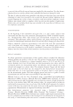
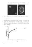
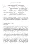
53 POTENTIAL BLUE LIGHT EFFECTS ON AGING Liebel’s study on the effect of visible light on ROS continued with human subjects. The foreheads of volunteers were exposed to visible light 400–700 nm at 50 J/cm2, and ROS was measured by a hydrogen peroxide-sensitive fluorescent probe. Free radical production was shown to increase over the baseline by 85.5% (2). Falcone performed a study to measure effects of 453 nm blue light on healthy skin perturbed using tape stripping. Included were 22 healthy volunteers with a mean age of 21.9 and Fitzpatrick skin types I, II, or III. No toiletries were used 24 h prior, and no sunbathing was permitted 2 weeks prior to experiment. Histamine iontophoresis and tape stripping was performed on two separate spots 4 cm apart on the volar forearm. Irradiance was delivered in pulsed (PW) or continuous (CW) mode. 15 cm of perturbed area of volar forearm was then irradiated with blue light at 453 nm with a fluence of 18 J/cm2 at 10 mW/ cm2 for 30 min. The pulsed mode delivered the same fluence but included a peak irradiance of 200 mW/cm2. Skin surface temperature was also taken from thermocouples of either side of the irradiance area. Six tests were performed over the course of 2 weeks (16). Skin reaction measured by transepidermal water loss (TEWL), skin surface biomarkers interleukin (IL)-1α, IL-1RA, human beta-defensin (hBD)-1, and hBD-2 were measured by means of transdermal analyses patch, and reflectance confocal microscopy was used to measure SC and epidermal thickness. All biomarkers IL-1α IL-1RA, ratio IL-1RA/IL-1α, hBD-1, and hBD-2 increased 24 h after tape stripping during control week. All biomarkers, except IL-1a, increased during irradiation week in the group that was perturbed and irradiated compared to nonperturbed and nonirradiated group. The authors conclude that blue light 453 nm light at 18 J/cm2 results in an inhibitory effect on IL-1a, which will lead to a reduction in release of lamellar bodies, as IL-1a promotes lipid and lamellar body synthesis. This difference was not statistically significant when perturbed and the irradiated group was compared to perturbed nonirradiated group. At 24 h, TEWL was increased, which the authors suggest is due to the reduced IL-1a levels after irradiation and is the first step in the inflammatory cascade, typically increased following barrier disruption (16). At 72 h, no difference in TEWL and epidermal thickness was noted in comparison of irradiated group and control group, suggesting no antiproliferative effect of single treatment. Reactive oxygen species. ROS can cause damage to lipids and proteins and can signal release of inflammatory cytokines. Nakashima performed an in vivo study looking at red, blue, and UV light using reduction-oxidation sensitive green fluorescent protein (roGFP), a fluorescent protein which can be oxidized by glutathione disulfide (GSSG), which is the oxidized form of cellular glutathione, a major redox protection system in human cells (3). If roGFP is present in the cells, it indicates that glutathione is responding to oxidative stress in the cells. Generation of oxidative species results in damage to skin lipids (17) and signal release of inflammatory cytokines by keratinocytes. roGFP was used due to its change in absorption upon oxidation, so quantification was simplified. Two species of mice were used—one expressed the roGFP in mitochondria, and the other expressed the roGFP in the cytosol and nucleus. Human subjects were also used and experiments performed were conducted within the parameters of the Declaration of Helsinki. The hand of participants was irradiated for 10 min at wavelengths of 400–480 nm at fluences of 11 mW/cm2. Blue light caused oxidative response via glutathione cascade in mitochondrial roGFP1 cells, as found by autofluorescence measurements, but not cytosol-based roGFP1 cells as shown in the mouse study.

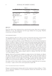
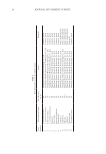
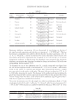
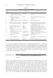
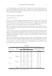
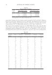
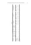
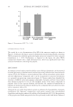
54 JOURNAL OF COSMETIC SCIENCE Since the oxidation rate tapers off at higher fluences, the authors conclude that endogenous flavin chromophores are destroyed by blue light. This flavin resulted in autofluorescence and was subtracted from mouse studies to isolate roGFP measurements. Human mitochondrial versus cytosolic oxidative cell responses mirrored mouse experimental results. Wavelengths in the green light region had no effect on oxidative events in the cytosol or the mitochondria. It was found that blue light cannot produce singlet oxygen as found by analysis of NADH and NADPH fluorescence measurements. Human skin exposed to blue light showed a decrease in autofluorescence measurements. Oxidation and conversion of cellular glutathione to GSSG does not occur very often in healthy cells usually roGFP will remain in a reduced state. This means that if skin cells are under stress conditions, there may be more GSSG existence in the cells. This chain reaction will occur as a result of superoxide and singlet oxygen. The author concludes that blue light was confirmed to contribute to skin aging by a slow and steady exposure to ROS, not by overwhelming cellular antioxidant levels by acute exposure. Melanin and carotenoids both absorb blue light as well as UVA light, but not green light, suggesting that evolutionarily, they are needed to protect the skin from blue light damage as well as UVA. Human skin exposed to normal sunlight levels of blue light showed a disintegration of endogenous flavoproteins (3). As discussed in earlier sections, this can lead to ROS which can signal activation of the MAPK/ERK pathway. Although the glutathione redox state was activated, no long-term blue light oxidative stress is indicated by this specific study as this is a short-term event. Per photon efficacy to activate Table II Blue Light-Induced Events in Comparison with Known UVA and UVB Events Blue light UVA UVB Penetration level Epidermis, dermis Epidermis, dermis Epidermis Main chromophores Flavins, heme, more data needed Flavins, heme, porphyrins, cytochromes Aromatic amino acids, nucleic acids Inflammatory cytokines & inflammatory cytokine-induced MMP IL-1a, MMP1, MMP9, reduction in TNF-alpha, no effect on IL-8 IL-1a, IL-6, IL-8, TNF-alpha TNF-alpha, MMP1, MMP9, MMP3 ROS species known No O2, NO, H2O2, more data needed O2, OH, NO, H2O2 O2, OH, NO, H2O2 DNA impact 8-oxoguanine, T4 endonuclease V (hamster cells in vitro only), more data needed 8-hydroxyguanine and formamidopyrimidines, T4 endonuclease V Pyrimidine dimers Keratinocytes Decrease in viability due to differentiation Decrease in viability Decrease in viability Fibroblasts Reduction in viability for wavelengths below 453 nm Reduction in viability Reduction in viability Wrinkles May lead to wrinkles by loss in dermal integrity Known to lead to wrinkles Known to lead to wrinkles Melanogenesis Through OPN3, may lead to age spots via combination of loss of dermal integrity and melanogenesis in skin types III and above Through p53, known to lead to age spots Through p53, known to lead to age spots
Purchased for the exclusive use of nofirst nolast (unknown)
From: SCC Media Library & Resource Center (library.scconline.org)