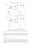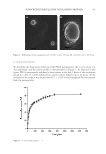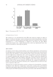
51 POTENTIAL BLUE LIGHT EFFECTS ON AGING activates melanogenesis enzymes, tyrosinase, and dopachrome tautomerase (DCT) (13). Levels of MITF, actin (which aids in melanin transport), DCT, and tyrosinase were tested in irradiated cells against controls immediately as well as 3 d following irradiation, which indicate melanogenesis activity. Actin and MITF were shown immediately after irradiation tyrosinase, DCT, actin, and MITF were present 3 days after irradiation. Reactive oxygen species. NRF2 is a transcription factor that responds to cellular stress such as reactive oxygen species (ROS) and UV light. Its accumulation indicates stressful conditions. It can signal antioxidant response systems to neutralize ROS and other harmful elements. It signals the upregulation of phase 2 antioxidant responders (e.g., glutathione, heme oxygenase, and peroxiredoxins) and UVA light is known to activate NRF2. In a study using human epidermal carcinoma cells injected into mouse skin irradiated with blue light from 400–500 nm, NRF2 was shown to be activated post- irradiation with blue light. Mitochondrial activity decreased beginning at a fluence of 15 J/cm2 and had an 80% reduction at 45 J/cm2 after 72 h of exposure (14). This indicates that there is an increase in ROS activity, and therefore a response in antioxidant systems in the skin. Oxidation events in the skin can contribute to damage in lipid and protein structure, leading to decreased tissue integrity. Inflammatory cytokines. Liebel studied normal human neonatal keratinocytes to analyze free radical production induced by visible light 400–700 nm at 65–180 J/cm2. Liebel exposed these keratinocytes to TNF a , UV (290–400 nm), and either 65, 130, or 180 J/cm2 of visible light, and tested the cells for markers of MAPK/ERK using Western blot. Increased phosphorylation of the epidermal growth factor, detected as phosphor-tyrosine, and the downstream marker of proliferation p42/44 MAPK was detected in the cells treated with TNF a , UV light, and visible light. This suggests activation of the MAPK/ERK pathway (2). Pretreatment of the cells with Tyrphostin, an EGFR inhibitor, blocked the downstream effects on ERK. This suggests that MAPK/ERK resulted from the activation of EGFR. The author concludes that visible light can induce MAPK/ERK in keratinocytes similarly to UV light. There was an increase in ROS formation, IL-1a, MMP1, and MMP9 levels, and in the presence of TNFa, an increase of markers of MAPK/ERK pathway activation. However, these levels only reached the levels of UVA/UVB when exposed directly to levels that exceed those delivered by normal sunlight (2). Liebel’s study on free radical production induced by visible light 400–700 nm at 65–180 J/cm2 was continued using human epidermal equivalents. Cytokine levels were calculated from testing the maintenance medium of the human epidermal equivalents 24 h post-irradiation by immunoassay multiplex kits. The level of MMPs and ROS became significantly increased beginning at 65 J/cm2, while the level of IL-1a became significantly increased beginning at 130 J/cm2 (2). IN VIVO STUDIES Nitric oxide. NO is a signaling molecule in human skin that can also lead to vasodilation. In addition, NO combines with ROS to form peroxynitrite, which is capable of producing lipid peroxidation and damage (15). This can deplete endogenous antioxidant systems in the skin and lead to damage of moisture-enhancing skin lipids, leading to decreased moisture content. In humans, enzymatic NO is produced by dermal cells. Mitochondrial respiration
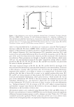
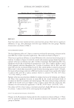
52 JOURNAL OF COSMETIC SCIENCE can be downregulated by NO (13). Some of the NO molecules formed remain close to their origin as nitroso compounds (e.g., S-nitrosothiols), mercuric chloride nonsensitive nitroso compounds, or the oxidation products nitrite and nitrate. It is known that these derivatives can begin decomposing when exposed to UVA to form bioactive NO. Several studies looked at whether blue light could affect NO release. Liebmann used keratinocytes and epithelial cell cultures in vivo, and irradiated them at 412, 419, and 426 nm at 66–100 J/cm2, and at 453 nm at 500 J/cm2. (12) As shown in Liebmann’s study, adenosine triphosphate levels also increased in keratinocytes and epithelial cells at 453 nm 500 J/cm2 (12). At longer wavelengths of blue light, such as 470 nm, mitochondrial respiration is reactivated. Liebmann estimates that this increase in adenosine triphosphate level is due to the release of NO from NO-sensitive complexes of the respiratory chain, such as bovine serum albumin, which act as photo acceptors, as a result of longer wavelengths of blue light. Liebmann used neonatal keratinocytes as well as reconstituted human epidermal tissue to study the effects of blue light on ROS and cytokines. These cells were exposed to differing doses of blue light ranging from 412–453 nm, UVA at 30 J/cm2, 412 nm at 33 J/cm2, 426 nm at 66 J/cm2, and 453 nm at 100 J/cm2. Blue light was found to have no effect on IL-8 release of human keratinocytes and endothelial cells (12). However, this study also showed that keratinocytes exposed to UVA also did not show an upregulation of IL-8 levels only in endothelial cells did this increase for UVA. IL-8 release can lead to a positive feedback loop and upregulation of elastase, leading to dermal matrix breakdown (15). This study suggests that neither blue light nor UVA light contributes to extrinsic aging from the single cytokine IL-8. For Opländer’s in vivo studies, volunteers were irradiated with a blue light dose of 30 J/ cm2 for 15 min. Opländer also used a closed chamber to collect NO gas from the forearm of human volunteers. An irradiation of 453 nm produced a small increase in gaseous NO emanating from the skin. Wavelengths in the green and red region did not produce a difference in NO levels. 420 nm, 52 J/cm2 produced a slight increase in S-nitroso-protein in homogenates of human skin. Cutaneous blood flow was analyzed with a micro-lightguide spectrophotometer. This resulted in greater NO formation than in vitro studies of human keratinocytes and resulted in NO-induced changes such as dilation of arteries and decrease in blood flow (10,11). There is a copper-dependence to the NO formation. A solution of nitrite-containing 10 mM NaNO 2 , as well as bivalent copper ions 2 mM CuCl 2 , were irradiated with blue light emitting LEDs (420 nm, 453 nm, 50 mW/cm2). Only the copper solution produced NO and did not produce NO with addition of a copper chelator. NO production was linear as a function of irradiance at 453 nm, and NO production peaked at 420 nm (10,11). Inflammatory cytokines. Kleinpenning studied the buttocks skin of eight females of Fitzpatrick skin types I–III with an average age of 20.9, via biopsies following exposure to 20 J/cm2 blue light at 420 nm. The skin was tested for p53, deformed elastin, MMP1, hyperpigmentation, inflammatory cells, keratinocytes, and sunburn cells. MMP1 was found to be present in five volunteer slides but was not significantly different than nonirradiated slides. The level of inflammatory cytokines also did not show a significant increase in irradiated slides from the controls. The author specified that, although blue light does not appear to contribute to extrinsic aging from acute exposure, no conclusions could be drawn from this study about the long-term effects of blue light exposure by the results of this study (19).
Purchased for the exclusive use of nofirst nolast (unknown)
From: SCC Media Library & Resource Center (library.scconline.org)