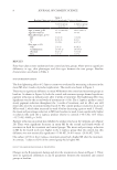
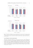
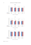
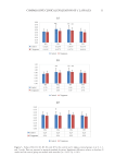
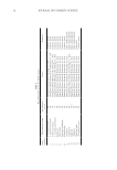
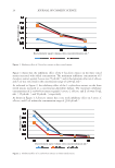
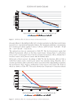
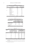
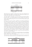
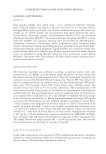
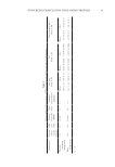
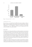
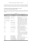
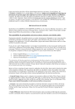
55 POTENTIAL BLUE LIGHT EFFECTS ON AGING the glutathione redox state is about 25% of that of UVA light. It would have been beneficial to repeat this study using times of exposure that were more long term (3). This study indicates that there is short-term oxidative events that occur as a result of blue light exposure. COMPARISON OF BLUE LIGHT SKIN INTERACTIONS WITH UVA AND UVB LIGHT SKIN INTERACTIONS Table II summarizes the differences between the interactions of skin with the different levels of light, as well as the tissue-level effects of the light-activated mechanisms that have been examined. Table III Summary of Previously Examined Data of the Mechanisms Activated by Blue Light Wavelength Fluence Result 410, 412, 415, 419, 420, 426, 453, 460, 470 nm 11 J/cm2 [Nakashima] Reduction in endogenous flavoproteins in vivo 410, 412, 415, 419, 420, 426, 453, 460, 470 nm 50 J/cm2 [Liebel] Hydrogen peroxide generated from human foreheads in vivo 410, 412, 415, 419, 420, 426, 453, 460, 470 nm 65 J/cm2 [Liebel] Increased ROS, no increase in IL-1a, increase in MMP1 and MMP9, Activation of MAPK pathway when in the presence of TNFa 410, 412, 415, 419, 420, 426, 453, 460, 470 nm 130 J/cm2-180 J/cm2 [Liebel] Increased ROS, increase in IL-1a, increase in MMP1 and MMP9, Activation of MAPK pathway when in the presence of TNFa 410 nm 60 J/cm2 [Oplander] Significant reduction in fibroblast viability 412 nm 33 J/cm2 [Liebmann] No effect on IL-8, reduction in keratinocytes and endothelial cells due to differentiation 415 nm 50 J/cm2 [Regazzetti] OPN3 dependent calcium flux causes upregulation of melanogenesis in FSTs III and above 419 nm 33 J/cm2 [Liebmann] Reduction in endothelial cells and keratinocytes due to differentiation 420 nm 60 J/cm2 [Oplander] Significant reduction in fibroblast viability 420 nm 20 J/cm2 [Kleinpenning] No impact on MMP, No impact on inflammatory cytokines, No increase on p53 levels, which increase melanogenesis 426 nm 66 J/cm2 [Liebmann] No effect on IL-8, no reduction in keratinocytes, reduction in endothelial viability 453 nm 18 J/cm2 [Falcone] Decreased IL-1a levels in epidermis following perturbation, increase in TEWL following perturbation, short-lived increase in blood flow following irradiation and perturbation, increase in melanin 72 h after irradiation and perturbation 453 nm 30 J/cm2 [Oplander] NO release in vivo 453 nm 90 J/cm2 [Oplander] No reduction in fibroblast viability 453 nm 100 J/cm2 [Liebmann] No effect on IL-8 460 nm 44 J/cm2 [Nakashima] Increases mitochondrial ROS in keratinocytes 470 nm 3 J/cm2 [Masson-Meyers] No reduction in fibroblast viability 470 nm 55 J/cm2 [Masson-Meyers] No reduction in fibroblast viability 470 nm 110 J/cm2 [Masson-Meyers] Reduction in fibroblast viability 470 nm 220 J/cm2 [Masson-Meyers] Reduction in fibroblast viability

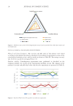
56 JOURNAL OF COSMETIC SCIENCE CONCLUSION Blue light has been shown to negatively impact the viability of fibroblasts (10,11,18). Acute exposure to blue light does not have any impact on elastin levels (19). However, by way of MMP1 and MMP9, this leads to the breakdown of both collagen and normally oriented elastin in the dermal layer, resulting in long-term contribution to extrinsic aging, with a loss in ability to resist deformation as well as the ability to recover from applied stress. At the epidermal layer, blue light caused melanogenesis in Fitzpatrick skin types III and higher (13). For individuals with this skin type, this can mirror age-spotting extrinsic aging events if the increase in melanogenesis is combined with a loss in dermal integrity (8,9). Blue light can also increase keratinocyte differentiation activity, which leads to a reduction in proliferation. This causes the epidermal turnover rate to decrease, and the stratum corneum will thicken. Kleinpenning reported a reduction in inflammatory cells however, this study did not give any specifics regarding the types of inflammatory cells that were examined (19). To contradict Kleinpenning, Liebel’s study did report an increase in ROS and IL-1 a levels (2). This research should be expanded to find specifics on how different inflammatory markers can be affected by certain wavelengths of blue light. Table III shows a summary of the previously examined experiments at differing wavelengths and fluences. Wavelengths below 420 nm are likely to contribute to extrinsic aging based on the elicited cellular mechanisms at these wavelengths, while wavelengths 453 and higher are relatively safe at sunlight-level fluences. REFERENCES (1) J. Moan, 7 visible light and UV radiation. Radiation at home, outdoors and in the workplace, Mater. Sci., (2004). (2) F. Liebel, S. Kaur, E. Ruvolo, N. Kollias, and M. D. Southall, Irradiation of skin with nonultraviolet light induces reactive oxygen species and matrix degrading enzymes, J. Am. Acad. Dermatol., 62(3) (2010). (3) Y. Nakashima, S. Ohta, and A. M. Wolf, Blue light-induced oxidative stress in live skin, Free Radic. Biol. Med., 108, 300–310 (2017). (4) T. C. Lei, S. Pendyala, L. Scherrer, B. Li, G. F. Glazner, and Z. Huang, Optical profiles of cathode ray tube and liquid crystal display monitors: implication in cutaneous phototoxicity in photodynamic therapy, Appl. Opt., 52(12), 2711–2717 (2013). (5) N. A. Monteiro-Riviere, Toxicology of the skin. Hoboken: Taylor & Francis (2013). (6) T. H. Quan, T. Y. He, J. J. Voorhees, and G. J. Fisher, Ultraviolet irradiation induces Smad7 via induction of transcription factor AP-1 in human skin fibroblasts, J. Biol. Chem., 280(9), 8079–8085 (2005). (7) M. Yaar, and B. A. Gilchrest, Ageing and photoageing of keratinocytes and melanocytes, Clin. Exp. Dermatol., 26(7), 583–591 (2001). (8) J. W. Choi, S. H. Kwon, C. H. Huh, K. C. Park, and S. W. Youn, The influences of skin visco-elasticity, hydration level and aging on the formation of wrinkles: a comprehensive and objective approach, Skin Res. Technol., 19(1), e349–e355 (2013). (9) W. Choi, L. Yin, C. Smuda, J. Batzer, V. J. Hearing, and L. Kolbe, Molecular and histological characterization of age spots, Exp. Dermatol., 26(3), 242–248 (2017). (10) C. Opländer, A. Deck, C. M. Volkmar, M. Kirsch, J. Liebmann, M. Born, F. A. van Abeelen, E. E. van Faassen, K. D. Kröncke, J. Windolf, and C. V. Suschek, Mechanism and biological relevance of blue-light (420–453 nm)-induced nonenzymatic nitric oxide generation from photolabile nitric oxide derivates in human skin in vitro and in vivo, Free Radic. Biol. Med., 65, 1363–1377 (2013).
Purchased for the exclusive use of nofirst nolast (unknown)
From: SCC Media Library & Resource Center (library.scconline.org)