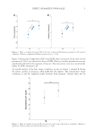
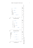
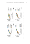
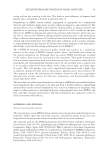
38 JOURNAL OF COSMETIC SCIENCE ANTIOXIDANT ACTIVITY DPPH–free radical scavenging. The scavenging activity of bee honey and AgNPs–bee honey nanoparticles samples were measured by using DPPH assay, and BHT was used as positive control. The unpaired electron of DPPH forms a pair with a hydrogen donated by free radical scavenging antioxidant from bee honey and AgNPs–bee honey nanoparticles, thus converting the purple colored odd electron DPPH to its reduced form of yellow (13). The degree of decolorization would be measured by UV-visible spectrophotometer to determine the scavenging activity of all bee honey samples. It was found that the percentage of radical scavenging activity of BHT was higher than all bee honey samples. The percentage of radical scavenging activity of bee honey samples is lower than AgNPs–bee honey nanoparticles samples because it requires a lesser amount of radical scavenger from the bee honey to reduce DPPH. The DPPH assays confirmed significant antioxidant activity of green synthesized AgNPs, which indicates the direct role of secondary metabolites, namely, phenolic compounds, terpenoids, and so on in removing exposed radicals. DPPH radical solution, which showed a deep purple color with maximum absorbance at 517 nm, turns into yellow color when it accepts an electron. Discoloration of DPPH started after adding AgNPs–bee honey nanoparticles due to the antioxidant ability radical scavenging activity of the bee honey sample increased with concentration from 125 to 500 μg/ml in the percentage of inhibition ranging from 43% to 95% (Figure 5). The AgNPs–bee honey nanoparticles exhibited the highest percentage of radical scavenging activity of 45% even with the lowest concentration of 125 μg/ml. Nevertheless, the lowest concentration of the AgNPs–bee honey nanoparticles (125 μg/ml) exhibited the highest percentage of radical scavenging activity, which is similar to that of the 125 μg/ml bee honey sample. Nanoparticles showed excellent antioxidant activity compared with that of the bee honey sample or other medicinal plants (14). Among tested samples, the strongest reducing antioxidant power measured by the FRAP test was found to be higher for AgNPs–bee honey nanoparticles than other bee honey samples (Figure 6). Figure 5. DPPH–free radical scavenging assay of bee honey samples (A, B, and C were bee honey from longan flowers, wildflowers, and benjaphan flowers, respectively) and the AgNPs–bee honey nanoparticles (a, b, and c were bee honey nanoparticles from longan flowers, wildflowers, and benjaphan flowers, respectively) at different concentrations demonstrated the percentage of radical scavenging activity.

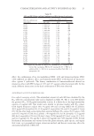
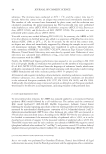
39 Silver Nanoparticles of Bee Honey ANTIMICROBIAL ACTIVITY In the present study, AgNPs–bee honey and bee honey were tested against S aureus and E coli. The zone of inhibition in diameter was discerned by disk diffusion method. For antibacterial function testing, S aureus, as well as E coli were used as the bacilli. The clear zone diameter of the bacterial inhibition zone was correlated to antibiotic activity (oxacillin and gentamicin). The clear zone diameter increased as the concentration of AgNPs–bee honey increased because of the bactericidal activity of Ag (15). The AgNPs–bee honey showed higher activity against gram-positive than gram-negative. The clear zones diameter of the bacterial inhibition zone for S aureus and E coli at a concentration of 1,000 µg/ml were 5.0±0.2 mm and 3.0±0.5 mm, respectively. AgNPs–bee honey exhibited effective zone of inhibition against S aureus and E coli more than the bee honey sample. However, AgNPs– bee honey showed better activities compared to bee honey. The highest zone of inhibition was observed for S aureus even at high concentration (≥250 µg/ml). The mechanism of the inhibition of the bacteria is still unknown, but some hypothetical mechanisms show that the inhibition is due to ionic binding of the silver nanoparticles on the surface of the bacteria, which creates a great intensity of the proton motive force, and the one hypothesis from the research states that the silver nanoparticles invade the bacterial cell and bind to the vital enzymes containing thiol groups (16). SERUM FORMULATION The F1-nano serum formulation containing 2% bee honey and AgNPs–bee honey nanoparticles was selected as best due to good physical properties such as homogeneity and fluidity, which certainly would make the serum application easier. The stability test of serum formulation was investigated. The obtained serums were submitted for 3 months and taken to accelerated stability study by heating/cooling for 3 cycles (kept at 45°C for 1 month and 4°C for 1 month/1 cycle) according to previous reports (17). The serums were then characterized by pH value and viscosity (18). The results showed that the formulations of F0-nano and F1-nano were not changed visually. These serums had a brawny and smooth texture with a bee honey odor. Thus, these formulations were used to evaluate characteristics Figure 6. Ferric reducing antioxidant power assay of bee honey samples and the AgNPs–bee honey nanoparticles at different concentrations.
Purchased for the exclusive use of nofirst nolast (unknown)
From: SCC Media Library & Resource Center (library.scconline.org)