REGIONAL VARIATIONS IN ENZYMES OF THE EPIDERMIS By RICHARD A. ELLIS, PH.D* Presented September 15-16, 1960, Seminar, Chicago THE CHEMICAL COMPOSITION of the epidermis has been examined by biologists and biochemists in some detail (7, 11, 23). Many of the organic and inorganic substances found in epidermal cells have been identified and studied under normal, experimental and pathological conditions. Since the epidermis consists of a highly heterogeneous population of cells, the biochemist has been largely unsuccessful in correlating specific chemical substances with a particular cell type. Using special techniques, some investigators have been successful in separating the keratinized cells from the rest of the epidermis, and analyzing the specific activities of these two epidermal components (10). In general, however, the biochemist has not been very successful in localizing specific substances within each of the lower layers of the epidermis. Within the limits of his methods, the histo- chemist has been more successful in this respect. Specific enzymes have been demonstrated in each of the lower layers of the epidermis, and it is primarily with the regional variability in the concentration of these en- zymes in the epidermis that this paper is concerned. Students of morphology have described the kaleidoscopic patterns that they have seen in comparing the structure of the epidermis from one part of the body with that from another. Variation in the thickness of the epidermis is apparent to anyone who has never contrasted a histological section of the palm with one of the eyelid for example. This variability in epidermal thickness is characteristic of many of the higher primates as well as man (20, 21, 25). More subtle differences in thickness are evident throughout the entire epidermis. The epidermis is about 27 microns thick over the roedial surface of the leg, 32 microns in the sacral region, 35 microns thick in the scapular region and 42 microns thick over the knee. Discrete differences also exist in the relative thickness of the various layers that comprise the epidermis. The stratum corneum may be three times as thick as the Malpighian layer in one locus, while at another site the Mal- pighian layer may be three times the thickness of the corneum. The stratum lucidurn may be poorly differentiated in one place, or 50/• thick in * Dept. ooe Biology, Brown University, Providence 12, P,. I. 64

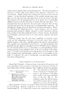
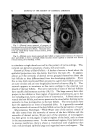
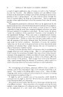
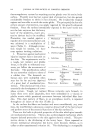
VARIATIONS IN ENZYMES OF THE EPIDERMIS 65 another the granulosum may vary in a similar manner. Although the basal layer, by definition, is only one cell thick, its surface area may be greatly increased in regions where the rete pegs are well developed. There are also measurable differences in the distribution of melanin and carotinold pigments in the epidermis (6) but these will not be considered in this paper. The appearance of the outer surface of the epidermis also varies remarkably over the body. In some regions sulci and ridges are present, in other places there are lines of flexure and folds. Since dermatoglyphics, lines of flexure and folds on the outer surface of the skin appear during prenatal life they probably have a genetic basis (22.27). These epidermal features also show distinct regional variation. Dermatoglyphics are present only at specific sites on the digits, pahns and soles. Lines and folds are also found at characteristic sites. The wrinkles and folds associ- ated with ageing have characteristic regional patterns, too. Less con- spicuous but even more dramatic is the complex sculpturing of the under surface of the epidermis (11, 16). In some regions the derreal-epidermal junction is almost flat in others there is a simple or complex system of fete ridges of varying sizes that interdigitate with the subjacent papillary body of the dermis. The character of sculpturing on the under surfaces determines the area of the basal epidermal layer that is available for meta- bolic exchange as well as the number of basal cells which may participate in mitosis. It is apparent, then, that the epidermis is not everywhere the same. It is also likely that the epidermal patterns are under genetic control, and that environment, except where it produces obvious or pathologic effects, does not influence greatly the normal structure of the epidermis. At the present time, however, we do not really understand how the genetic ma- terial produces the spectrum of epidermal patterns that have just been described. It is the purpose of this paper to present some recent work that contrib- utes to our understanding of the basis for the regional differences that are seen in the epidermis, and to discuss just how such studies may be able to explain the observed variability in the epidermal patterns. It is not improbable that such information may lead us to the factors which govern the development of normal as well as abnormal epidermal structure. Using histochemical techniques for demonstrating alkaline phosphatase activity (13, 17), the smaller blood vessels supplying the epidermis can be visualized with remarkable clarity. This is possible because the en- dothelial cells lining the venules, capillaries and arterioles contain sub- stantial quantities of alkaline phosphatase. This method is superior to injection or perfusion procedures, since even collapsed vessels may be demonstrated. In skin specimens taken from various sites on the same subject, striking
Purchased for the exclusive use of nofirst nolast (unknown)
From: SCC Media Library & Resource Center (library.scconline.org)