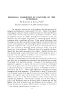
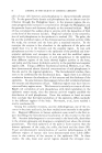
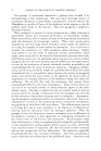
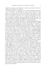
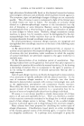
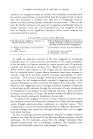
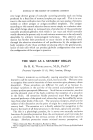
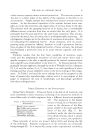
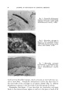
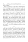
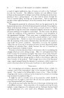
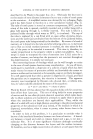
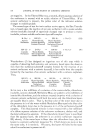
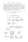
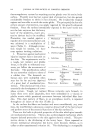
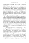
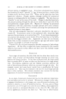
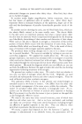
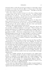
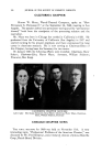
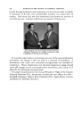
82 JOURNAL OF THE SOCIETY OF COSMETIC CHEMISTS Fig. l.--Dermal nerve network of prepuce of human newborn, demonstrating meshwork of nerves containing associated nerve fibers. Granular struc- tures are small blood vessels (frozen section, silver method X160). Fig. •2.--Multiple nerve trunks approach hair follicle at region below entrance of sebaceous duct and divide to form a network which becomes closely applied to external root sheath (frozen section, silver method X760). to stimulate a single dorsal-root cell in this pattern of nerve endings. This network can perceive sensations of pain, itch and touch. Network of Nerves of Hair Fo//ic/e.--A basket of nerves is found about the epithelial projections into the dermis that form the hairs (9). It appears almost as if the networks of derreal nerves grouped themselves about the hair follicles as they differentiated from the developing epidermis. From five to nine thick myelinated fibers proceed to a given hair follicle and form an outer circular and an inner longitudinal network of nerves (Fig. 2). This network appears to terminate as free nerve endings in the external root sheath of the hair follicle. The nerve networks of some of the hair follicles have specific cholinesterase activity (10, 11). The large sensory hairs that project in the whisker or brow region of mammals and primates, up to but not including man, have a special form of innervation. A blood sinus sur- rounds the hair follicle, and nerves penetrate the sinus before forming nerve networks in close juxtaposition to the hair follicle. The terminations here have the appearance at times of expanded disks. It is generally conceded that the network of nerves of the hair follicle serves the perception of touch. Mucocutaneous End-Organ.--While 90 per cent of the sensory stimuli of the skin comes from the networks of nerves of the hair follicles and the networks of derreal nerves, 90 per cent of the attention of investigators has been given to end-organs in special regions of the bod3 , such as those in the mucocutaneous zones. In the transitional regions, from haired skin to mucous membrane, there is always a transitional zone of glabrous skin with well-developed epithelial pegs and organized nerve end-organs. The

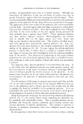
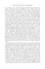
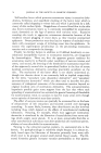
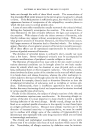
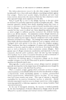
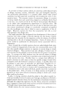
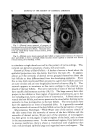
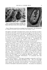
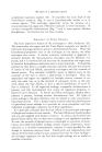
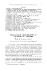
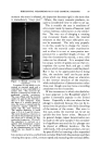
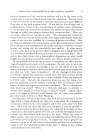
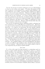
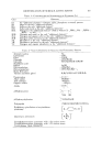
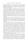
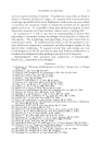
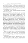
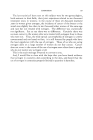
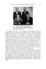
THE SKIN AS A SENSORY ORGAN 83 Fig. &--Typical mucocutaneous end-organ com- posed of rolled, nonmyelinated fibers, from prepuce of human adult (frozen section, silver method X 670). Fig. 4.--Meissner's corpuscle from volar digital skin of human being. Note expanded disk- like ending and multilobular shape (frozen section, silver method X 265). end-organs in the region of the glans pe.nis, clitoris, lip, conjunctiva and peri- anal skin are all similar, and for this type of end-organ we use the term "mu- cocutaneous end-organ," for it indicates the morphologic unit that exists in the nervous tissue of all these regions (12). The mucocutaneous end-organ is composed of a ball of rolled nonmyelinated nerve fibers which proceed from three to four myelinated nerve trunks (Fig. 3). Nonspecific cho- linesterase activity is present (13). While it is not definitely known, it is assumed that this end-organ supplies the sensation of acute touch. Meissner's Corpusc/e.--In that part of the skin of the hands and feet in primates where the surface is friction bearing, a variation of the muco- cutaneous end-organ exists that has been termed "Meissner's corpuscle" (Fig. 4). This end-organ terminates in expanded disks or networks and has such a distinctive structure that it may be recognized by simple tissue stains such as hematoxylin and eosin. An aborization of tactile disks may also be found along the base of the rete ridges of the epithelium of fingers and toes. This is occasionally seen in regions such as the lip of the cat. These end-organs are supplied by clusters of nerve fibers of a thick mye- linated type and appear to be unorganized Meissner corpuscles. They contain pseudo or nonspecific cholinesterase. It is presumed that they serve the sensation of touch. lZater-Pacini Corpuscle.--The largest end-organ in mammals is the'Vater- Pacini corpuscle, which was first described in 1741. This is an end-organ that is not found in the skin alone but appears also near joints, in the pro- static capsule, the mesentery, and the pancreas. It is found in animals as diverse as the chicken and the boa constrictor, It consists of a concentric,
Purchased for the exclusive use of nofirst nolast (unknown)
From: SCC Media Library & Resource Center (library.scconline.org)