PATTERNS OF REACTION OF THE SKIN TO INJURY 71 (26) (27) REFEKENCES (1) Argyris, T. S., ?[nat. Record, 125, 105 (1956). (2) Bejdl, W., Z. Zellforsch. u. mikroskop. ?[nat., 40, 389 (1954). (3) Braun-Falco, O., bfaut ?[rch. klin. exptl. Dermatol., 202, 153 (1955). (4) Bullough, W. S., Biol. Rev., 27, 133 (1952). (5) Bullough, W. S., and Lawrence, E. B., "The Mitotic Activity of the Follicle," in "The Biology of Hair Growth," W. Montagna and R. A. Ellis, Editors, New York, Academic Press, Inc. (1958), pp. 171-172. (6) Edwards, E. A., and Duntley, S. Q., ?ira. y. ?[nat., 65, 1 (1939). (7) Ellis, R. A., Ageing of the Human Male Scalp, in "The Biology of Hair Growth," W. Montagna and R. A. Ellis, editors, New York, Academic Press, Inc. (1958), pp. 469- 485. (8) Ellis, R. A., and Montagna, W., y. Histochem. and Cytochem., 6, 201 (1958). (9) Herrmann, F., Harbev, L. C., Scher, R., and Mondol, L., ?[.M.?[. ?[rch. Dermatol., 76, 282 (1957). (10) Hershey, F. B., Lewis, C., Murphy, J., and Schiff, T., y. Histochem. and Cytochem., 8, 41 (1960). (11) Horstmann, E., Die Haut, in "Handbuch der mikroskopischen Anatomie des Menschen," W. v. Mollendorff, Ed., $ (1), (1957), pp. 1-276. (12) Katzberg, A., ?[nat. Record, 112, 418 (1952). (13) Kopf, Alfred W., ?[.M.?[. ?[rch. DermatoL, 75, 1 (1957). (14) Loeb, L. and Haven, P. L., ?[nat. Record, 42, 217 (1929). (15) Montagna, W., 5 t. Biophys. and Blochem. CytoL, 1, 13 (1955). (16) Montagna, W., ]bid., $, 343 (1957). (17) Montagna, W., and Ellis, R. A., y. Natl. CancerInst., 19, 451 (1957). (18) Montagna, W., and Formisano, V., ?[nat. Record, 122, 65 (1955). (19) Moretti, G., Adachi, K., and Ellis, R. A., y. Histochem. and Cytochem., 8, 237 (1960). (20) Moretti, G., Adachi, K., Mescon, H., and Pochi, P., in press (1961). (21) Moretti, G., Ellis, R. A., and Mescon, H., y. Invest. Dermatol., 33, 103 (1959). (22) Rawles, M. E., The Skin and Its Derivatives, in "Analysis of Development," Willier, Weiss and Hamburger, editors, Philadelphia, W. B. Saunders Co. (1955), pp. 499-519. (23) Rothman, S., "The Physiology and Biochemistry of Skin," Chicago, Univ. of Chicago Press (1954). (24) Schering, L. E., ?[nat. Record, 135, 7 (1959). (25) Straus, W. L., Jr., The Microscopic Anatomy of the Skin of the Gorilla, in "The Anatomy of the Gorilla," W. K. Gregory, ed., New York, Columbia Univ. Press (1950), pp. 213- 226. Thuringer, J. M,., and Katzberg, A. A., y. Invest. Dermatol., 33, 35 (1959). Montagna, W., 'The Structure and Function of Skin," New York, Academic Press, Inc. (1956). PATTERN S O F REACTION O F THE SKIN TO INJURY By ALL^N L. Lo}ttNcz, M.D.* Presented September 1546, 1960, Seminar, Chicago A•ao• T•E PA??ER•S of reaction of the skin to injury two general ones which may be elicited by a wide variety of factors are encountered so frequently as to deserve special consideration as rather fundamental ele- ments in cutaneous pathology. These reaction patterns are namely ec- zematous and urticarial. The eczematous reaction pattern has greater significance for the cosmetic chemist because it is so very often triggered by substances coming into contact with the skin. * Assdciate Professor of Dermatology, The University of Chicago, Chicago, Ill.

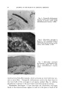
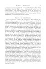
72 JOURNAL OF THE SOCIETY OF COSMETIC CHEMISTS The concept of eczematous dermatitis is perhaps best clarified if its histopathology is first understood. The most basic histologic feature of eczematous dermatitis is intercellular accumulation of serous fluid in the Malpighian or prickle-cell layer of the epidermis which appears to be the primary shock tissue in the reaction. The term spongiosis is applied to this type of edema. When spongiosis is minimal it can be recognized by a slight widening of intercellular spaces and increased prominence of intercellular bridges. When more severe, there is rupture of some of the intercellular connections with the formation of microscopic vesicles. When most severe, gross vesiculation and even large blister formation occurs within the epidermis as is seen, for example, in acute poison ivy dermatitis. It is of interest to consider the mechanism by which spongiotic edema develops. Clearly such edema is not the result of enhanced vascular permeability which might cause serous fluid to be forced by hemodynamic pressure from leaky small blood vessels into the epidermal spaces, because one cannot produce spongiosis by even the most forceful injection of fluid into the upper dermis or even by the production of greatly enhanced vascular permeability and vessel leakage there by means of histamine injections. Spongiosis, further- more, cannot be accounted for simply by a possible shift of fluid from inside of epidermal cells to intercellular spaces because the volume of spongiotic edema may exceed the total volume of the epidermis by many orders of magnitude, the average thickness of the epidermis being only about 0.1 millimeter. We are left then with the assumption that osmotic forces essentially pull fluid into the areas of spongiosis. These osmotic forces can act only if an increased number of solute molecules appear in the inter- cellular spaces. Damage to epidermal cells can readily result in such an increase partly by leakage of substances from the damaged cells, and also by the activation of proteolytic enzymes such as cathepsins. Accompany- ing the spongiotic epidermal reaction in eczematous dermatitis there is an inflammatory reaction in the underlying corium marked by vasodilation, the gathering of white blood cells, and edema. This inflammatory reaction is, in large part and at least, secondary to the liberation of inflammation exciting humoral substances from the damaged epidermis. With the passage of time the eczematous reaction undergoes some modification as the epidermis attempts to heal apparently by undergoing acceleration of the normal rates of cell division and maturation. Keratinization under these circumstances is often incomplete both physiologically and chemically so that parakeratotic scaling appears which consists of softer, chemically more reactive keratin in squamous cells that still retain nuclear remnants and are less reorganized and less dehydrated. The epidermis, especially the prickle-cell layer may increase in thickness. The term, acanthosis, is used to describe such thickening of the prickle-cell layer. Acanthosis and
Purchased for the exclusive use of nofirst nolast (unknown)
From: SCC Media Library & Resource Center (library.scconline.org)