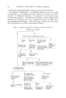
PATTERNS OF REACTION OF THE SKIN TO INJURY 77 reactions into categories based on whether the antibodies associated with the reaction remain fixed or closely linked with the lymphold cells in which they were produced or whether they spill over i'n biologically reactive amounts as free antibody protein molecules into the serum. Thus, as the basis for further discussion the generally recognized morphologic types of allergic reactions of the skin can be grouped into two categories on this basis of whether or not significant quantities of free serum antibody are associated with the reaction. CLASSIFICATION OF ALLERGIC SKIN REACTIONS With Biologically Significant Amounts of Specific Antibody in Serum Without Biologically Significant Amounts of Specific Antibody in Serum but with Such Antibody Associated with Lymphocytes (1) Allerg. ic anglo-edema and urticarial re- actions (2) Arthus reaction (1) Eczematous contact type skin sensitivity (2) Tuberculin type sensitivity (3) Homograft immunity As might be expected, reactions in the skin triggered by circulating antibodies have (1) a short reaction time because of the ready availability of the antibody in all places reached by serum protein, and (2) primarily vascular and perivascular reaction sites. Reactions thus, usually occur within minutes after exposure to the antigen and are characterized in the skin by urticarial wheal or giant edema formation based upon damage to vascular walls with attendant greatly increased permeability of these structures. This vascular damage is believed to result in fairly large meas- ure at least by the antigen-antibody reaction's triggering action on the release of histamine as well as serotonin and heparin from mast cells. Fur- thermore, this triggering action of antigen-antibody complexes is believed to be at least partly mediated through the activation of some components of complement to proteolytic or ester splitting enzymes. Such activated proteases and esterases, of course, can produce additional effects by means other than simply causing the breakdown of mast cells. In general the antigens involved in these urticarial reactions reach the cutaneous vessels via the systemic circulation to which they have gained access through gastro-intestinal, respiratory or injection routes. Very exceptionally, in cases of extreme hypersensitivity, sufficient absorption of a protein antigen may occur even through unbroken skin so as to trigger local wheal reactions. In man, circulating antibody encountered clinically in allergic urticarial reactions can often be detected by the Prausnitz-Kustner passive transfer technique. In this technique, serum from the allergic individual when injected intradermally into a nonallergic recipient's skin produces tem- porary local urticarial reactivity to the antigen in question.

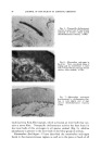
78 JOURNAL OF THE SOCIETY OF COSMETIC CHEMISTS The Artbus phenomenon occurs in the skin when antigen is introduced intradermally into a host with much effective circulating antibody against that antigen. Severe local vascular injury, thrombosis and even local tissue necrosis characterize this reaction which has been observed to occur after repeated foreign serum injections into the skin. Next, I would like to turn to the allergic reactions in the skin which are mediated by antibodies apparently carried on lymphocytic cells. These reactions generally develop more slowly requiring twenty-four hours or more or sometimes even as long as a week to become manifest after contact with the allergen. Furthermore, the reaction site is not so primarily peri- vascular but may occur in almost any tissue depending apparently simply on where antigen in sufficient quantity encounters the antibody bearing cells to set off the reaction. In eczematous contact allergic reactions, as exemplified by poison ivy dermatitis, the eliciting substances are commonly relatively simple reactive organic chemical compounds such as the catechol derivative involved in ivy allergy. On coming into contact with the skin, these simple eliciting compounds are believed to act as happens which must conjugate with some protein in the skin, to form the complete antigens. Thus, conditions that favor conjugation of various such compounds with proteins in the skin would be expected to be factors favoring the develop- ment as well as elicitation of these eczematous contact allergies. Such greater ease of allergic eczematous sensitization and reactivity in fact are commonly observed under conditions where the skin has been damaged with resultant loss of some of its barrier functions as well as formation of incompletely keratinized chemically more reactive keratin. Sometimes the eliciting agents of eczematous allergy can be already complete antigens as can be well illustrated by positive eczematous contact reactions to tuberculin patch tests. Histologically, in acute eczematous contact allergic reactions spongiosis generally develops first in the deeper layers of the epidermis even though the eliciting agent comes to the skin from the surface. When a complete antigen, against which antibodies carried on lympho- cytes are present, is introduced locally into the skin intradermally or deeper, a characteristic, slowly developing, papular or nodular, circumscribed chronic granulomatous, inflammatory reaction develops. If very severe, even local tissue necrosis may occur. This type of reaction is classically exemplified by the positive intradermal reaction to tuberculin. Similar delayed type papular reactions occur in allergic sensitivity to other antigens occurring especially in infectious organisms such as viruses, bacteria, fungi and animal parasites. In the case of homograft immunity, the reaction is marked primarily by the slow development of chronic inflammatory changes about the graft followed by vascular damage and eventual necrosis of the grafted tissue.
Purchased for the exclusive use of nofirst nolast (unknown)
From: SCC Media Library & Resource Center (library.scconline.org)