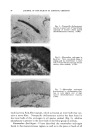
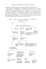
PATTERNS OF REACTION OF THE SKIN TO INJURY 75 leaks out through the walls of these blood vessels. The accumulation of this transuded fluid under pressure in the dermis gives rise grossly to wheals or hives. If the fluid pressure is sufficiently great, the wheal has a blanched appearance because of compression of the subpapillary vascular plexus to which the skin owes its normal pinkish color. , A zone of axon reflex vasodilation usually surrounds the urticarial wheal. Itching also frequently accompanies the reaction. The amount of hista- mine liberated in the skin critically influences the signs and symptoms of the reaction. With release of only the tiniest amounts of histamine, only blotchy redness may appear without accompanying itching. With some- what greater amounts of histamine liberation, red wheal formation occurs. Still larger amounts of histamine lead to blanched wheals. For itching to appear, liberation of even greater amounts of histamine is usually necessary. All of these effects can be reproduced experimentally by intradermal in- j•ctions of graded amounts of histamine. The duration of urticarial lesions is ordinarily short, usually not more than a few hours. Severe widespread urticaria may be accompanied by systemic manifestations of peripheral vascular collapse or shock. The liberation of histamine from mast cells in the skin under normal or various abnormal conditions can be brought about by two basic mecha- nisms by stimuli which may be chemical or even physical such as cold, heat, mechanical forces or electromagnetic radiation. One of these mecha- nisms involves the direct action of the stimulus on the mast cell which causes it to break down and release histamine, whereas the other mechanism in- volves injury to the mast cell brought about by the indirect route of allergy. A whiplash for example, directly leads to histamine liberation as does the intradermal introduction of substances such as morphine or various ami- dines. Unfortunately, there is insufficient opportunity here to consider further the many fascinating clinical and experimental intricacies involved in various specific types of urticaria. Finally in this discussion, the subject of allergic reactions of the skin and their mechanisms requires some consideration. In discussing allergic re- actions of the skin it is most important to keep clearly in mind a specific limiting definition of the word allergy. Briefly stated, allergy can be de- fined as an acquired specific alteration in the capacity to react based upon an antigen antibody reaction. Because most allergic reactions are in the direction of increased reactivity or hypersensitivity, we must be careful not to confuse with allergy some of the varieties of hypersensitivity or idiosyncrasy seen especially with some drug reactions which are not based on immunologic mechanisms. The clinical and pathological manifestations of allergy can be quite diverse even if we consider only those of such reactions which occur in the skin. Nevertheless, all allergic reactions by definition must be immuno-

76 JOURNAL OF THE SOCIETY OF COSMETIC CHEMISTS logic phenomena fundamentally based on the chemical interaction between some antigenic substance and antibodies which had been formed against it. The symptoms, signs and pathologic changes of allergy are not necessarily specific. This, of course, is easy to understand in light of the limited ways in which tissues can react to injury. For example, an urticarial wheal elicited as a pharmacophysiologic response to the introduction into the. skin of histamine or some histamine liberator cannot be distinguished morphologically from a wheal arising as a specific manifestation of allergy to some antigen in lobster meat. Similarly, allergic eczematous contact reactions to poison ivy, for example, cannot be distinguished by the der- matopathologist from similar reactions that can be elicited by primarily irritating chemicals through nonallergic mechanisms. For practical purposes, cutaneous allergic reactions are recognized and distinguished from nonallergic reactions on the basis of the following types of evidence: (1) By demonstration of specific skin hypersensitivity or unusual re- activity on test exposure to the suspected allergenic material, often by means of common skin test procedures, under conditions in which the majority of normal individuals not previously exposed to the agent would fail to similarly react. (2) By demonstration of an incubation period of sensitization. Sup- porting evidence here can be gained (a) from proof that prior exposure to the material in question did not elicit the unusual reactivity, (b) by showing a shortening of reaction time on re-exposure to the agent in question, or (c) by noting the phenomenon of delayed spontaneous flare-up after a single exposure. And (3) such allergic reactions are finally distinguished by demonstration of an association of specific antibodies with the altered reactivity. In the case of cutaneous allergic reactions it is relatively rare that direct laboratory demonstration succeeds with common immunologic tests such as the agglutination, precipitation and complement fixation reactions. More often, less direct means are necessary for this demonstration such as passive transfer of the allergic sensitivity to a nonsensitive host by in- jections of serum or lymphold cells taken from the allergic individual. Of course, it is not always possible to clearly demonstrate all of these points in all cases and especially the last. There are, therefore, a number of skin reactions commonly considered to be allergic but in which complete proof of their allergic nature is lacking. Many of the so-called allergic reactions to physical agents and to drugs belong in this uncertain category. Many schemes have been proposed for classifying the various types of allergic phenomena. Classifications, based purely on either morphology of the reaction, general nature of the eliciting agent, or reaction time are not altogether satisfactory. It is perhaps more meaningful to group the
Purchased for the exclusive use of nofirst nolast (unknown)
From: SCC Media Library & Resource Center (library.scconline.org)