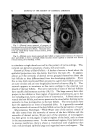
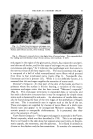
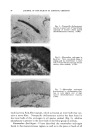
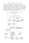
66 JOURNAL OF THE SOCIETY OF COSMETIC CHEMISTS differences are apparent in the form, number and distribution of blood vessels supplying the epidermis. Where the epidermis is thin, and where the fete pegs are poorly developed, the capillary loops are few and the vascular bed scant. In the thigh, for example, and in the temporal region, the blood vessels supplying the epidermis are few in number and archi- tecturally simple. In the perioral region and in the scalp, the capillaries form complete loops that arch within each derreal papilla. In the pressure regions such as the palm and the sole, the circulatory pattern is most elab- orate, and is consistent with the complex character of the dermal-epidermal junction. One might conclude that there is a direct correlation between the thick- ness of the epidermis and the richness of its blood supply. This, however, is not always true. For example, on the forehead the epidermis is 45 to 65/a thick and there are an average of 157 capillary loops per min. 2, while on the nose, the epidermis is 75/a thick and it has an average of 100 under- lying capillary loops per min. 2 (21). In neither region are the fete pegs well developed. In general, however, the development of the fete ridges can be correlated with a rich vascular bed. This is shown dramatically in the ageing changes that take place in the scalp (7). In the child and in the young male, the epidermal pegs of the scalp are pronounced and long capil- lary loops course through each of the interdigitating derreal papillae. With increasing age, the derreal-epidermal junction is flattened, and the loops are lower and less numerous. In the bald scalp the epidermis is nearly fiat along its base, and there are only a few low-lying, arched capillaries beneath. It is not surprising that the vascular architecture in the papillary body of the dermis is often associated with variations in the structure of the epidermis. It is unfortunate, however, that we are unable to separate cause and effect in this case. Does the vascular pattern in the papillary body of the dermis influence epidermal structure, or does the epidermis affect the vascular pattern? At present we cannot answer this question, and indeed, the answer to each may be affirmative. Under normal con- ditions, all of the metabolites consumed by the epidermis are carriedJto it through the walls of the underlying blood vessels. The greater the number of vessels, the larger the quantity of nutrients that can be supplied. If the turnover of the population of epidermal cells is dependent on its supply of nutrients, one would expect a greater mitotic rate as well as a higher metabolic rate in regions with a rich blood supply. The scant information available on the mitotic rate and the replacement rate of epidermal cells indicates that there are distinct regional differences in each of these in- dices (12, 14, 23, 24, 26). Since enzymatic activities are frequently a reflection of the metabolic activity of a tissue, close examination of the enzymes present in the epidermis is of paramount importance. Recent

VARIATIONS IN ENZYMES OF THE EPIDERMIS 67 biological evidence suggests that enzymes are under the close control of the genetic material of the cell. Thus, studying variability in the activity of the epidermal enzymes may indicate how the genes influence cellular differentiation. Variation in the concentration and localization of histochemically demon- strable enzymes in the epidermis has been recognized for several years. Usually these differences were noted in regions where the structure of the epidermis was markedly different. The localization of nonspecific a-naph- thol esterase in the epidermis of the palm and sole showed a strong en- zymatic activity in the lower cells of the Malpighian layer, but almost none in the layers above, while in the thinner epidermis of the axilla most of this enzyme was concentrated in a band between the stratum comeurn and the stratum granulosum (15). It was suggested that in the epidermis esterase hydrolyzed lipids during the process of keratinization, thus splitting off free fatty acids. The striking differences observed in distribution of esterase within the epidermis from different regions of the body was not explained. The epidermal activity of other esterases has been demonstrated histo- chemically. Indoxyl acetate esterase is present in all but the horny layer of the epidermis (3). Possible regional differences in the activity of this enzyme have not yet been examined. Tween 60 esterase has been localized in human epidermis as well as in the epidermis of other primates (19, 20). This method demonstrates both lipases and esterases. Tween 60 esterase has its greatest concentration in the Malpighian layer of the epidermis. With semiquantitative methods, this enzyme has been found to vary sig- nificantly in epidermis from various regions of the body. In the chim- panzee the enzyme is richest in the brow and the sole, and least reactive in the Malpighian layer of the axilla and the lower lip. In the macaque epidermal activity is strongest in the scrotum and in the forehead and weakest in chest and in the nape. The data indicate that the concentration is two to three times greater at sites of high activity than it is in regions giving a low reaction. The concentration of this enzyme does not seem to correlate with any of the morphological differences observed in the epidermis. Argyris (1) has studied changes in the esterase content of the epidermis of the mouse, and reports that enzyme is strongest during the resting phase of the hair growing cycle, and that the esterase activity de- creases when the hair of the mouse is in the growing phase of the cycle. In primates it is unlikely that esterase activity is correlated with hair growth, since the cyclic activity of the hair follicles does not sweep through the epidermis in predictable waves as it does in the mouse (27). Neither does the esterase activity appear to correlate with the distribution of hair nor with the length of hairs on the body surface. There is very little or no alkaline phosphatase in any of the epidermal
Purchased for the exclusive use of nofirst nolast (unknown)
From: SCC Media Library & Resource Center (library.scconline.org)