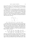
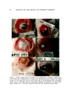
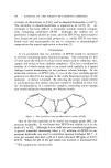
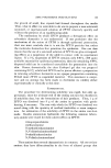
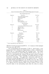
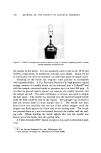
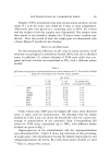
94 JOURNAL OF THE SOCIETY OF COSMETIC CHEMISTS conjunctival sacs the left eyes were injected intracorneally with 0.01 ml (7 X 10⁴ cells) of a suspension of pseudomonas organisms. Four pseudo- monas-treated incised rabbit eyes were used as positive controls. Re- sults of this ,test, though exploratory, strongly suggest that monkey eyes are not as seriously affected by these pseudomonas organisms as are rabbit eyes. All 4 rabbit eyes showed typical pseudomonas keratitis, ending in perforation in 10 days. All 4 monkey eyes showed effects which were less severe than those in rabbit eyes. The maximum effects (in all monkey eyes) occurred in 48-72 hours and consisted of a white corneal opaci. ty with redness of the lids. This persisted for several weeks with gradual improvement but incomplete resolution (Fig. 1). Experiments on Tissues Other Than the Cornea A series of experiments was conducted to investigate •the susceptibility of tissues other than the cornea to invasion from pseudomonas organisms or injury from its products. Both tissues investigated, the sclera and the skin, are vascular tissues, in contrast to the cornea. In addition, their collagens may be different biochemically. Studies on Sclera--Incisions about 5 mm long were made with a scalpel deep into the scleral fibers of three rabbits. Immediately thereafter, 0.1 ml (7.5 million cells) of a suspension of pseudomonas organisms was in- stilled into the conjunctival sacs. Findings were essentially negative. Only slight injection of blood vessels was observed at the incision sites. Intrascleral injection of pseudomonas organisms, however, produced more dramatic effects than those seen above. The right eyes of each of 4 rabbits were injected with 0.02 ml (3 X 10⁵ cells) of a suspension contain- ing pseudomonas organisms. The left eyes (positive con•trols) were ad- ministered about 1.5 X 10 ø cells in the conjunctival sacs after the corneas were incised with a scalpel as described previously. Intrascleral adminis- tration produced upper lid redness and chemosis and a central abscess surrounded by injected blood vessels. Peak effects were reached in 1-3 days with gradual resolution in about 2-3 weeks (Fig. 1). The posi,tive controls all developed'typical pseudomonas keratitis. Studies on Skin--A straight incision about 5 cm long was made with a scalpel deep into the dermis on the clipped backs of 12 animals. Each cut was filled with a suspension containing about 7.5 million pseudo- toohas organisms. Examination of these sites at various times thereafter showed no significant skin reactions. Intradermal injection (0.1 ml/site) of the backs of 2 rabbits with a suspension containing 15 million pseudornonas •cells per site resulted in

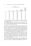
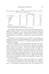
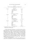
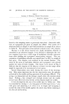
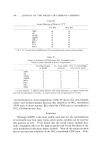
PSEUDOMONAS KERATITIS 95 erythema and edema (about 2.5 cm in diameter) in 24 hours. At 48 hours, a 1-cm area ot• necrosis was observed it persisted for at least 7 days. Similar results were obtained by injecting 2 additional rabbits with 1.5 million cells per site except that the areas ot• erythema, edema, and ne- crosis were reduced. Intradermal injection with 0.15 million cells re- sulted in only slight edema in two other rabbits. Intradermal injection ot• 0.1 ml ot• pseudomonas endotoxin produced erythema and edema in 24 hours, but no subsequent necrosis, as dis- tinguished from the effect with pseudomonas organisms. Mechanism o[ Tissue Destruction in Pseudomonas Keratitis A number of investigators have suggested that the tissue-destructive propensities ot• P. aeruginosa reside in the capacity ot• the organism to elaborate a collagenolytic enzyme resembling collagenase (9, 10). In the next series of experiments, we were able to demonstrate that this was in- deed the case. Furthermore, this enzyme appears to require activation in vivo. Corneal buttons were excised t•rom rabbits 72 hours after intracorneal injection with P. aeruginosa. (Pseudomonas endotoxin was used in simi- lar experiments.) TT• ,,• adequate positive and negative controls, these corneal tissues, which were undergoing destruction in vivo, were excised, incubated with suitable buffers for 24 hours, and tested for collagenolytic activity according to the method of Mandl et al. (11). Results, shown in Table II, demonstrate that corneas which are ac- tively undergoing tissue destruction by P. aeruginosa or by pseudomonas endotoxin are indeed capable of releasing amino acid breakdown prod- ucts of collagen when tested as described. No evidence of active col- lagenase w. as demonstrated in pseudomonas cultures alone or when such Table II Results of Tests for Collagenolytic Activity a Collagenase q- collagen q- Collagenase q- cornea q- Cornea, 72 hr after intracorneal injection of P. aeruginosa q- Cornea, 72 hr after intracorneal injection of pseudomonas endotoxin q- Pseudomonas culture -- Pseudomonas culture q- collagen -- Pseudomonas culture q- cornea -- Endotoxin -- Endotoxin q- cornea -- According to method of Mandl et al. (ll).
Purchased for the exclusive use of nofirst nolast (unknown)
From: SCC Media Library & Resource Center (library.scconline.org)