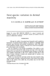
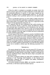
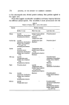
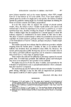
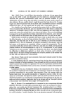
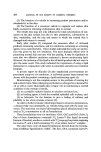
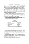
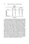
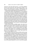
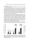
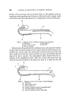
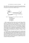
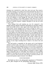
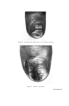
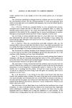
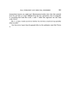
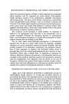
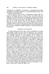
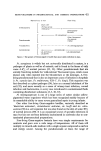
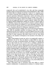
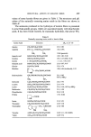
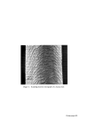
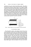
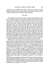
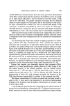
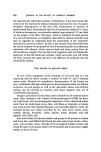
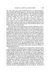
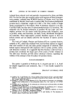
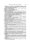
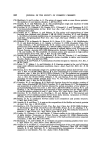
406 JOURNAL OF THE SOCIETY OF COSMETIC CHEMISTS portion of the roof may also be involved (Fig. 1). The portion of the in- vagination which produces nail is known as the matrix and is largely under cover of the roof of the nail fold and the overlying skin at the base of the nail. b e d Figure 1. Diagram to show traditional theory of nail formation: (a) Nail plate. (d) Nail matrix. (b) Skin at base of nail. (e) Nail bed. (c) Roof of nail fold. Barton Lewis (1) questioned the traditional view and produced evidence to show that the nail is formed in three layers which he called dorsal, inter- mediate and ventral nails. The dorsal nail is formed from part of the roof and a small part of the floor of the nail fold, but may be lost before the free edge of the nail is reached.. The intermediate nail is formed from the re- mainder of the traditional matrix, while the ventral nail arises from the whole of the nail bed distal to the half-moon (Fig. 2). A third and very old c b Figure 2. Diagram to show Lewis's theory of nail formation: (a) Nail plate. (b) Skin at base of nail. (c) Roof of nail fold. (d) Portion of matrix forming 'intermediate nail'. (e) Nail bed forming 'ventral nail'. (f) Portion of matrix forming 'dorsal nail'.

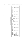
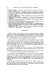
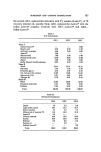
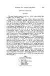
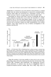
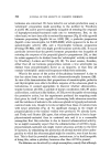
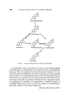
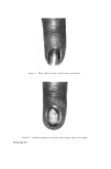
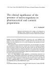
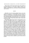
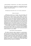
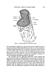
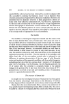
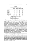
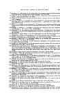
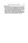
NAIL FORMATION AND SOME NAIL DISORDERS 407 theory (Boas (2)) maintains that the exposed nail bed is sterile except for a small portion close to the point of separation of the nail from its bed. This area is given the German name 'solenhorn' (Fig. $). Figure $. Diagram to show method of nail formation according to Boas: (a) Nail plate. (b) Skin at base of nail. (c) Roof of nail fold. (d) Nail matrix. (e) Sterile nail bed. (g) 'Solenhorn'. There is support for all three theories and the author believes all three methods of nail formation may occur. Support for the traditional view comes from experiments with the squirrel monkey, an animal with a flat nail very similar to that of man (3,4). Support for Lewis's view comes from histochemical staining (5) and from electron-microscopic studies (6). Large amounts from the nail bed are seen in a few pathological conditions. The third view would be supported by any surgeon who has undertaken total nail ablation only to find spicules of nail appearing later close to the tip of the digit. The mammalian claw is formed in two layers so that the alternative methods of nail formation may represent a reversion to a more primitive state. The rate of growth of finger nails in health varies from 1.2 mm per week down to 0.5 mm. There is a gradual slowing with age, so that most of the more rapidly growing nails will be found in children and the slower rates in old age. Pathological conditions can alter the rate of nail growth. The com- monest cause for increased rate of growth is psoriasis, but many nails when they become partially separated from their bed (onycholysis) also grow faster than normal. Very slow rates of growth are usually the result of a few uncommon developmental conditions.
Purchased for the exclusive use of nofirst nolast (unknown)
From: SCC Media Library & Resource Center (library.scconline.org)