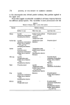
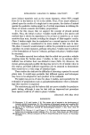
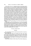
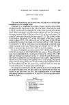
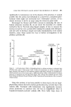
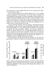
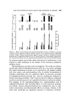
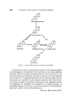
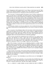
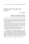
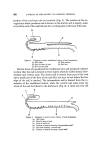
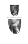
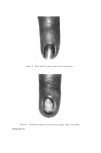
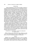
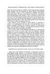
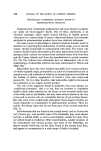
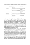
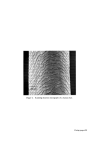
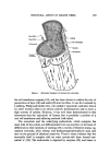
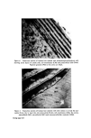
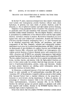
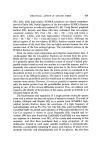
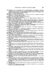
430 JOURNAL OF THE SOCIETY OF COSMETIC CHEMISTS of the aspartic acid residues in wool appear to be aminated (10, 11) and that the carboxyl groups are present mainly as glutamic acid residues (11). It can be seen that all keratin fibres contain a very high level of sulphur, most of which occurs as cystinc. Human hair has less alaninc, leucine, tyrosine, phenylalanine, glutamic acid, aspattic acid, lysine, and arginine than other keratin fibres and is richer in cystinc and proline. These differences probably indicate a higher proportion of high-sulphur proteins, as it is known that human hair has a greater extent of cross-linking than most wool fibres. The heavier cross-linking is reflected in greater resistance to attack by hot acids and in slower reduction by thioglycolate or sulphite solution (12). The ready uptake of dyes by mohair is probably due to the presence of a high content of ionizable side-chain groups (13). Human hair, on the other hand, has a lower content of the amino acids with ionic side-chains than mohair or wool this difference is reflected in its dyeing behaviour. Thus, human hair has a lower capacity for acids (14) as well as for acid dyes, e.g. Orange II (15), than wool. On the basis of dyeing and swelling experiments, some investigators have suggested that human hair is predominantly 'para' (vide infra) in its properties (16). KERATIN FIBRE HISTOLOGY Keratin fibres are very complex both at the histological level and at the chemical level owing to the multiplicity of protein molecules which are effectively cross-linked to form an integral structure. Histologically, keratin fibres consist of three main components: (a) the cuticle cells (about 10• of the fibre), which envelop the fibre and overlap rather like files on a roof (b) the cortical cells (about 88• of the fibre), which are long spindle-shaped cells aligned parallel to the fibre direction and (c) the cell membrane complex (about 2• of the fibre), which separates each cuticle or cortical cell from its neighbour. The medulla is present in some types of fibres it consists of a core of air-filled cells which runs down the middle of the fibre. Fig. 1 is a schematic diagram of human hair structure and Fig. 2 is a scanning electron micrograph of a human hair. The cuticle The outermost layer of the cuticle was observed by Allwbrden (17) as a membrane which was raised from the surface of wool by treatment with chlorine water. It surrounds each cuticle cell individually, forming part of

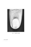
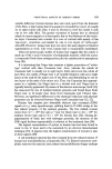
Figure 2. Scanning electron micrograph of a human hair. Facing page 430
Purchased for the exclusive use of nofirst nolast (unknown)
From: SCC Media Library & Resource Center (library.scconline.org)