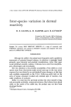
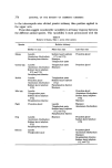
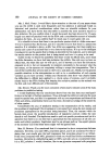
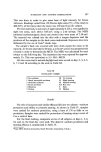
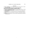
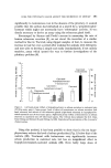
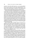
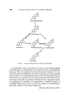
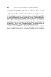
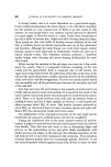
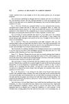
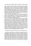
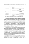
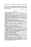
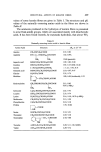
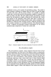
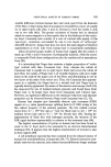
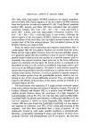
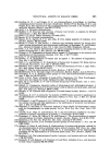
NAIL FORMATION AND SOME NAIL DISORDERS 407 theory (Boas (2)) maintains that the exposed nail bed is sterile except for a small portion close to the point of separation of the nail from its bed. This area is given the German name 'solenhorn' (Fig. $). Figure $. Diagram to show method of nail formation according to Boas: (a) Nail plate. (b) Skin at base of nail. (c) Roof of nail fold. (d) Nail matrix. (e) Sterile nail bed. (g) 'Solenhorn'. There is support for all three theories and the author believes all three methods of nail formation may occur. Support for the traditional view comes from experiments with the squirrel monkey, an animal with a flat nail very similar to that of man (3,4). Support for Lewis's view comes from histochemical staining (5) and from electron-microscopic studies (6). Large amounts from the nail bed are seen in a few pathological conditions. The third view would be supported by any surgeon who has undertaken total nail ablation only to find spicules of nail appearing later close to the tip of the digit. The mammalian claw is formed in two layers so that the alternative methods of nail formation may represent a reversion to a more primitive state. The rate of growth of finger nails in health varies from 1.2 mm per week down to 0.5 mm. There is a gradual slowing with age, so that most of the more rapidly growing nails will be found in children and the slower rates in old age. Pathological conditions can alter the rate of nail growth. The com- monest cause for increased rate of growth is psoriasis, but many nails when they become partially separated from their bed (onycholysis) also grow faster than normal. Very slow rates of growth are usually the result of a few uncommon developmental conditions.

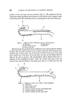
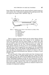
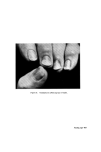
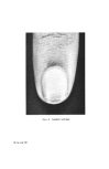
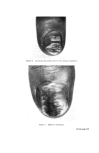
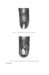
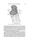
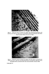
408 JOURNAL OF THE SOCIETY OF COSMETIC CHEMISTS A strong healthy nail is of course dependent on a good blood supply. Under normal circumstances the blood supply to the nail fold is abundant but the arteries are very exposed and are liable to go into spasm in cold weather. In some people this is very common and the nail may be deprived of a good supply of blood for hours at a time. Under these circumstances the nail is liable to become thin, ridged and splits develop along the ridges. These people are also very liable to develop chronic infection of the nail fold, a condition known as chronic paronychia and one of the commonest nail disorders. Although the same change can result from organic arterial damage, spasm is more important as anastomotic vessels can open up to replace injured vessels. Iron deficiency anaemia produces a somewhat similar change when thinning and spoon shaping (koilonychia) are most often found. Before leaving the anatomy of the nail organ one must say a few words about the cuticle. This is a compound structure consisting of the true cuticle and the eponychium. Each is composed only of soft keratin, the upper layer being derived from the epithelium of the skin at the base of the nail and the eponychium from a similar extension forward of the epithelium of the roof of the nail fold invagination. The two are usually closely united and successfully seal off the potential space between the nail plate and roof of the nail fold. Like hair and wool, the nail contains a fair quantity of water and it can readily take up more in moist surroundings. It is uncertain how much of the water content comes from deeper structures and how much from the atmos- phere. There is no doubt, however, that nails take up moisture freely on soaking in water and lose it fairly quickly on removal. A nail in good con- dition contains about 16% of water. Nail keratin becomes saturated at about 30% at this level it loses its lustre, becomes opaque and quite soft. Below 16}/o the nail becomes brittle and cracks easily. The implications of the anatomical structure of the nail organ and its interference by natural or artificial means will now be considered. Taking the traditional view as the most common method of nail for- mation, it might be expected that the nail would separate from the nail bed very readily. That this does not occur is due to alternating ridges and grooves on the undersurface of the nail plate which interdigitate with similar grooves and ridges on the nail bed. Nevertheless separation of the nail from its bed is one of the commonest nail symptoms. It is a symptom of many diseases mainly local but occasionally general. However, it can also occur frequently without apparent cause (Fig. 4) but in many of these
Purchased for the exclusive use of nofirst nolast (unknown)
From: SCC Media Library & Resource Center (library.scconline.org)