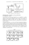
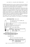
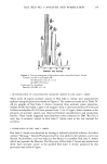
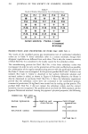
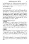
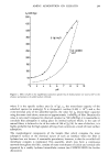
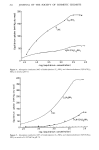
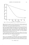
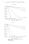
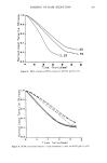
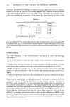
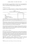
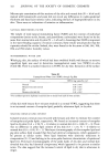
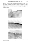
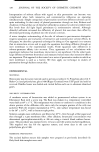
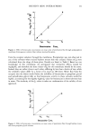
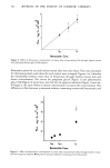
ANIMAL MODEL OF HUMAN DRY SKIN 319 tissue mounted in a ground-glass diffusion cell (14). Water-soluble materials were extracted from stratum comeurn as described by Blank (15). Amino acids (16-17) and lactate (18) were analyzed as described in the references cited. Pyrrolidone carboxylate was determined as glutamate (19) following heating at 100øC for 30 minutes in 3 N HCI and neutralization with NaOH (20). Stratum corneum hydration was done at room temperature over saturated solutions of salts at the indicated relative humidity (RH) values: CaCI 2 (30% RH), NaHSO4 (50% RH), NaCIO 3 (75% RH), KCI (85% RH), and K2SO4 (95% RH). For estimation of stratum comeurn lipid content, the stratum comeurn surface of whole skin was swabbed briefly with a hexane-saturated cotton-tipped applicator to remove surface lipid. The stratum comeurn was then isolated (12), dried, weighed, extracted with hexane for 24 hours with mild agitation, recovered by filtration (Whatman No. 1 filter paper), and reweighed. The tissue was then extracted with ether for 24 hours with mild agitation, filtered, and reweighed. The weight loss after hexane extraction and after ether extraction yielded the nonpolar and polar lipid contents, respectively. Thorough hexane extraction of the stratum corneum side of whole skin was done by clamping the tissue in a ground-glass diffusion cell with hexane in contact with the stratum comeurn for five minutes. In experiments involving sebum application to tissue, one mg of sebum per cm 2 was spread on the stratum comeurn. Sebum was obtained by swabbing the surface of pig or human skin with hexane, which was then filtered the hexane-soluble material (sebum) was recovered by removal of the solvent with a stream of dry nitrogen. Thin-layer chromatography of lipids was done by spotting a toluene solution of the lipids on 8-inch x 8-inch glass plates coated with silica gel G (Supelco Inc., Bellefonte, PA). The plates were developed with three consecutive solvent systems as follows: 1) chloroform:methanol:water at 90:10:1 (v:v:v) to half way up the plate, air dry 2) hexane:diethyl ether:acetic acid at 30:70:1 (v:v:v) to three-quarters way up the plate, air dry and 3) hexane:benzene at 3:2 (v:v) tO top of plate, air dry. The lipids were made visible by spraying the plate with 0.25% H2SO 4 and charting at 600øF for ten minutes. For experiments involving the generation of dry skin in vitro, whole normal skin was clamped in a ground-glass diffusion cell. The dermis side of the tissue was kept in contact with 0.9% NaCI, while dry nitrogen was blown across the stratum corneum surface. At timed intervals, tissue was removed and treated with sebum as described above. Whole human abdominal skin was obtained at autopsy. RESULTS AND CONCLUSIONS DEVELOPMENT OF DRY SKIN Housing of pigs in a low-relative-humidity (10%) environment resulted in development of dry skin (Figure 1), which was visually graded as indicated in Table I. Dry skin first appeared along the spinal area. The appearance of this dry skin was rapid, being noticeable to a significant degree in less than a day (Figure 2). The dry skin became generally distributed over the back within one to two weeks. The skin grade was nearly constant from shoulder to hip after housing the pigs for about one month, at which

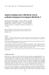
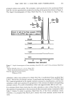
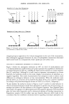
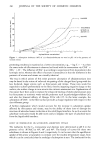
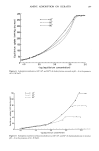
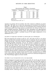
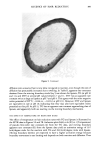
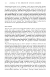
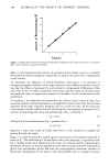
320 JOURNAL OF THE SOCIETY OF COSMETIC CHEMISTS , ' . Figure 1. Photographs of pig skin: A) normal skin (grade 0.5) and B) dry skin (grade 3.5). . 4 0 i i i i 2 4 6 8 Days at Low Relative Humidity Figure 2. Change in skin grade after placement of a pig with normal skin in a low-relative-humidity environment.
Purchased for the exclusive use of nofirst nolast (unknown)
From: SCC Media Library & Resource Center (library.scconline.org)