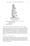
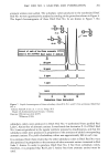
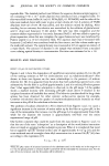
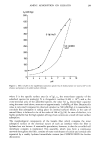
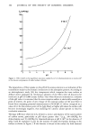
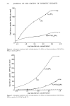
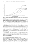
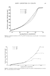
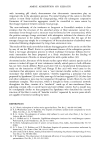
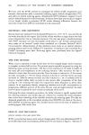
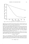
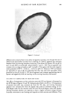
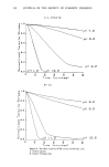
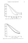
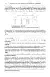
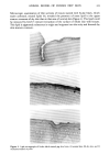
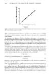
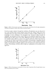
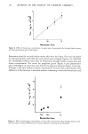
ANIMAL MODEL OF HUMAN DRY SKIN 325 this lipid entry may not occur in human dry skin to the extent that it does in pig dry skin. For dry and visually normal pig skin, no differences in stratum corneum thickness, turnover rate, water-binding capacity, or NMF content could be detected. There were also no differences observed in the viable epidermis. It must be noted, though, that the techniques used will not necessarily reveal subtle differences in the properties examined but certainly indicate the lack of gross differences. The rapid onset of dry skin observed here suggests the lack of involvement of the viable epidermis in the early stages of dry skin formation. However, there may be subtle biological changes in the viable epidermis as a response to the formation of dry skin in the stratum corneum. In humans, there is a variable susceptibility to dry skin formation among individuals under the same environmental conditions. There is certainly an underlying biological/ biochemical explanation for this variability, and a difference in the stratum corneum among individuals certainly exists, resulting in variable resistance to dehydration. The basis for the difference is not known. In pigs, we have not observed variable susceptibility to dry skin formation among individuals. All pigs tested developed dry skin. Anderson et al. (27) observed in humans increases in water-binding capacity, NMF content, and protein for occlusion-treated dry skin relative to untreated dry skin. However, as noted by the authors, the unrealistic total occlusion (from Saran © Wrap) probably increased skin temperature, resulting in an enhanced metabolic rate in the viable epidermis and an increased production of NMF and protein involved in water binding. We are most likely not obtaining total occlusion with petrolatum, and therefore would not encounter the differences reported by Anderson et al. Also, surfactant interactions with skin may be involved in the development of human dry skin. Comparative studies will be necessary to determine the degree of correlation between pig skin and human skin in their normal and dry states. The rapid rate of formation of dry skin on the pig at low relative humidity (Figure 2) suggests that this dry skin is a phenomenon of the stratum corneum and does not require the involvement of viable epidermis or any other insult such as surfactant exposure. Dry skin appears to involve only the upper portion of the stratum corneum (Figure 3). The presence of large surface scales and of sebaceous lipid deep within the stratum corneum indicates defective cell adhesion in dry skin. Since desquamation of surface cells from skin likely involves enzymatic processes (31-32), the aberrant cell adhesion in dry skin may be due to altered enzyme-catalyzed hydrolysis in the stratum corneum. REFERENCES (1) I. H. Blank, Factors which influence the water content of the sratum corneum,J. Invest. Dermatol., 18, 433-440 (1952). (2) P. Flesch and E. C.J. Esoda, Deficient water-binding in pathologic horny layers, J. Invest. Dermatol., 28, 5-13 (1957). (3) K. A. Grice and F. A. Bettley, Skin water loss and accidental hypothermia in psoriasis, ichthyosis, and erythroderma, Brit. Med. J., 4, 195-198 (1967). (4) P. Frost, G. D. Weinstein,J. W. Bothwell, and R. Wildnauer, Ichthyosiform dermatoses. III. Studies of transepidermal water loss, Arch. DermatoL, 98, 230-233 (1968).

326 JOURNAL OF THE SOCIETY OF COSMETIC CHEMISTS (5) C. Prottey, Essential fatty acids and the skin, Brit. J. Dermatol., 94, 579-587 (1976). (6) P.J. Hattop and C. Prottey, Changes in transepidermal water loss and the composition of epidermal lecithin after applications of pure fatty acid triglycerides to the skin of essential fatty acid-deficient rats, Brit. J. Dermatol., 95,255-264 (1976). (7) O. K.Jacobi, Moisture regulation in the skin, Drug. Cosmet. Ind., 84, 732-733, 810-812 (1959). (8) L. H. Jansen, M. T. Hojyo-Tomoko, and A.M. Kligman, Improved fluorescence staining technique for estimating turnover of the human stratum corneum, Brit. J. Dermatol., 90, 9-12 (1974). (9) J. F. A. McManus and R. W. Mowry, Staining Methods.' Histological and Histochemical (Paul B. Hoebert, New York, 1960), pp 110-111. (10) L. G. Luna, Manual of Histological Staining Methods of the Armed Forces Institute of Pathology (McGraw-Hill, New York, 1968), pp 36-39. (11) D. E. McOsker and I,. W. Beck, Characteristics of accommodated (hardened) skin,J. Invest. Dermatol., 48, 372-383 (1%7). (12) B. F. Van Duzee, Thermal analysis of human stratum corneum, J. Invest. Dermatol., 65, 404-408 (1975). (13) R. L. Rietschel, A method to evaluate skin moisturizers in vivo, J. Invest. Dermatol., 70, 152-155 (1978). (14) B. Idson, Percutaneous absorption,J_ Pharm. St'i, 64, 901-924 (1975). (15) I. H. Blank, Further observations on factors which influence the water content of the stratum corneum,J. Invest. Dermatol., 21,259-271 (1953). (16) S. Moore and W. H. Stein, Photometric ninhydrin method for use in the chromatography of amino acids,J. Biol. Chem., 176, 367-388 (1948). (17) S. Moore, Amino acid analysis: Aqueous dimethyl sulfoxide as solvent for the ninhydrin reaction, J. Biol. Chem., 243, 6281-6283 (1968). (18) H.J. Hohorst, Methods of Enzymatic Analysis (Academic Press, New York, 1965), pp 266-270. (19) E. Bernt and H. U. Bergmeyer, Methods of Enzymatic Analysis (Academic Press, New York, 1%5), pp 384-388. (20) B. Moav and T. N. Harris, Pyrrolid-2-one 5-carboxylic acid involvement in the biosynthesis of rabbit immunoglobulin, Biochem. Biophys. Res. Commun., 29, 773-776 (1967). (21) N. Nicolaides, H. C. Fu, and G. R. Rice, The skin surface lipids of man compared with those of eighteen species of animals,J. Invest. Dermatol., 51, 83-89 (1968). (22) G. M. Gray, R.J. White, R. H. Williams, and H.J. Yardley, Lipid composition of the superficial stratum comeurn of pig epidermis,, Brit. J. Dermatol., 106, 59-63 (1982). (23) W. Meyer, R. Schwarz, and K. Neurand, The skin of domestic mammals as a model for the human skin, with special reference to the domestic pig, Curr. Probl. Dermatol., 7, 39-52 (1978). (24) W. Montagna and J. S. Yun, The skin of the domestic pig,J. Invest. Dermatol., 43, 11-21 (1964). (25) G. D. Weinstein, Swine in Biomedical Research (Frayn, Seattle, 1966), pp 287-297. (26) G. M. Gray and H. J. Yardley, Lipid compositions of cells isolated from pig, human, and rat epidermis,J. Lipid Res., 16, 434-440 (1975). (27) R. L. Anderson,J. M. Cassidy,J. R. Hansen, and W. Yellin, The effect of in vivo occlusion on human stratum corneum hydration-dehydration in vitro, J. Invest. Dermatol., 61,375-379 (1973). (28) J. E. Kirk, Hand washing, Acta Dermato- Venereol., 46 (Suppl 57), 1-183 (1966). (29) E. Nieminen, E. Leikola, M. Koljonen, U. Kiistala, and K. K. Mustakallio, Quantitative analysis of epidermal lipids by thin-layer chromatography with special reference to seasonal and age variation, Acta Dermato- l/enereol., 47, 327-338 (1967). (30) M. Gloor, V. Willebrandt, G. Thomer, and W. Kupferschmid, Water content of the horny layer and skin surface lipids, Arch. Dermatol. Res., 268, 221-223 (1980). (31) A.M. Kligman, An overview of acne,J. Invest. Dermatol., 62,268-287 (1974). (32) P. C. Woo-Sam, Cohesion of horny cells during comedo formation, Brit. J. Dermatol., 97, 609-615 (1977).
Purchased for the exclusive use of nofirst nolast (unknown)
From: SCC Media Library & Resource Center (library.scconline.org)