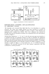
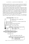
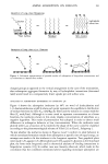
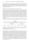
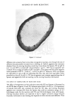
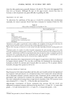
ANIMAL MODEL OF HUMAN DRY SKIN 323 Microscopic examination of thin sections of tissues stained with Sudan black, which stains unbound, neutral lipids (9), revealed the presence of more lipid in the upper stratum corneum of dry skin than in that area of normal skin (Figure 3). This lipid could be removed by brief (5 minute) extraction of the surface of whole skin with hexane. This lipid is apparently sebaceous in origin and migrates into the scaly and fissured dry skin stratum corneum. Figure 3. Light micrographs of Sudan black-stained pig skin from: A) normal skin, B) dry skin, and C) occlusion-treated dry skin.

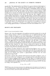
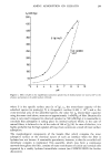
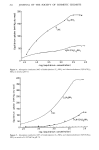
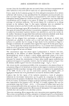
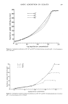
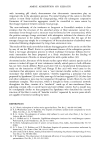
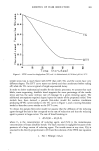
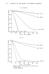
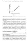
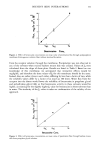
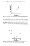
324 JOURNAL OF THE SOCIETY OF COSMETIC CHEMISTS Experiments to generate dry skin in vitro were done by passing a stream of dry nitrogen over the surface of whole normal pig skin. At timed intervals (0, 6, 24, 48, and 72 hours), the surface was treated with pig sebum, sectioned, stained with Sudan black, and examined microscopically. Slight staining of the stratum corneum could be seen at 6 hours, and by 24 hours the tissue was as darkly stained as in vivo-generated dry skin, with the staining confined to the outer half of the stratum corneum. Identical results were obtained at the 48- and 72-hour time points. Controls which were not exposed to dry nitrogen or which were not treated with sebum remained unstained, i.e., both drying and exposure to sebum were required for the observed staining. Identical results were obtained for human skin. While these tissues did not visually have the appearance of dry skin, cell cohesion is presumably being affected, as it is in vivo, to permit entry of sebum into the stratum corneum. These data and those in Figure 2 indicate that dry skin formation occurs rapidly under harsh environmental conditions. DISCUSSION There are many similarities between pig skin and human skin (21,23-26), including hair density, skin surface structure, epidermal structure, sebum composition, epidermal turnover time, and epidermal lipid composition. Added to the list now is the fact that pigs develop dry skin at low relative humidity this dry skin is visually like that observed on humans. The dry skin responds to treatment with known skin conditioners and to a return to high relative humidity conditions. This animal model is valuable as a tool for rapidly evaluating new skin conditioning actives. Particularly valuable is the ability to test several materials on the same animal, permitting direct comparisons of their efficacies. In contrast to the seasonal availability of human dry skin, pig dry skin can be obtained on a year-round basis with the appropriate controlled environmental facilities. This animal model is also useful as a tool to determine the differences between dry skin and normal skin. The only difference detected by the work done here was an increase in nonpolar lipid in the upper stratum corneum of dry skin. This was determined to be due to migration of surface sebaceous lipid into the cracks and fissures of dry skin, not to any actual change in the epidermal lipid composition. Anderson, Cassidy, Hansen, and Yellin (27) detected an increase in lipid (type undetermined) content of dry skin relative to occlusion-treated dry skin in humans. Also, sebum lipids (28), and triglyceride in particular (29), are elevated in human skin during the winter, the time when dry skin is most prevalent. For pig skin, this lipid difference resulted in a TEWL reduction for dry skin, a reduction which could be eliminated by extraction of the sebaceous lipid. In humans (30), no correlation has been seen between TEWL and skin surface lipid content. Presumably, this lipid is present as a thin film on the skin surface. In pig dry skin, the lipid is deep within the stratum corneum, forming a relatively thick film which could well provide a barrier sufficient to reduce TEWL. We do not believe that the increased sebum lipid content in pig dry skin is a causative factor in dry skin formation. The cracks and fissures of dry skin are simply providing a route for entry of lipid into the stratum corneum where it is retained rather than being lost with surface cells during desquamation. In our histological sections, there were no indications of sebaceous gland density or size differences in dry skin versus normal skin. Since humans routinely expose their skin to surfactant, removing a majority of sebum (28),
Purchased for the exclusive use of nofirst nolast (unknown)
From: SCC Media Library & Resource Center (library.scconline.org)