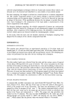
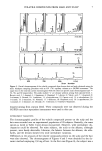
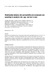
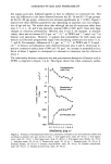
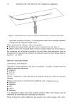
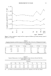
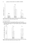
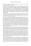
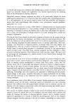
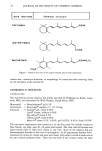
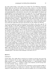
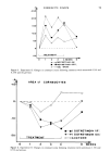
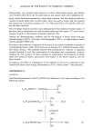
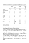
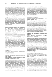
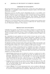
16 JOURNAL OF THE SOCIETY OF COSMETIC CHEMISTS on isolated corneocytes may provide important information concerning the function of the stratum corneum as a whole (11). In this paper we attempt to establish the relationship between the barrier properties of the horny layer (percutaneous absorption and TEWL) and the surface area of corneocytes according to anatomic site, age, and sex in man. MATERIALS AND METHODS PERCUTANEOUS ABSORPTION The penetration of benzoic acid was measured at seven anatomic sites, the location of which are depicted in Figure 1. A group of six to eight informed male volunteers, aged 20-30 years, was used for each anatomical site. The influence of aging on skin absorption of benzoic acid was studied on groups of seven to eight male volunteers, 45-55 and 65-80 years of age. The anatomic site involved was the upper-outer arm. The influence of sex was assessed on the upper-outer arm and on the forehead of groups of seven to eight female volunteers, 20-30 years of age. Application conditions. One thousand nanomoles of benzoic acid (ring-•4C) (New England Nuclear), of specific activity 10 -3 IxCi/nmol, were applied to an area of 1 cm 2 in 20 Ixl of a vehicle consisting of ethyleneglycol to which 10% Triton © X 100 had been added øG u Figure 1. Anatomic sites tested: A. Forehead. B. Postauricular. C. Arm (upper-outer). D. Forearm (ven- tral-elbow). E. Forearm (ventral-mid). F. Forearm (ventral-wrist). G. Abdomen.

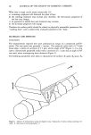
CORNEOCYTE SIZE AND PERMEABILITY 17 as surfactant. The treated area was demarcated by an open circular cell fixed by silicone glue to prevent chemical loss. After 30 minutes, excess chemical was quickly removed by two successive washes (2 X 300 }xl) with a 95-5 ethanol-water mixture, followed by two rinses (2 x 300 }xl) with distilled water and drying lightly with a cotton swab. Measurement conditions. Benzoic acid was selected on the basis of the speed and high level of its urinary excretion. Thus, utilizing literature data relating to the kinetics of urinary excretion of this compound administered intravenously and orally (12,13) or percuta- neously (14- 16) in different species, the percentage of the urinary excreted benzoic acid relative to that absorbed within the first 24 h was 75%. The total quantities absorbed during the four days following application could, therefore, be calculated, after scintil- lation counting, from the quantities found in the urine up to 24 h. TRANSEPIDERMAL WATER LOSS (TEWL) After completion of the benzoic acid treatment, TEWL was measured with an Evapo- rimeter EPIC (Servo Med, Sweden) from a contralateral site (same anatomic region) in each subject. The handheld probe was fitted with a 1-cm tail chimney to reduce air turbulence around the hydrosensors and metallic shield (supplied by Servo Med), mini- mizing the possibility of sensor contamination. Measurements (gm ß m -2 ß h-•) stabi- lized within 30-45 seconds. Since the room environment was comfortable (room tem- perature 20øC, relative humidity 70%) and the subjects physically inactive, the TEWL should closely reflect stratum corneum water flux without significant sweating interfer- ence. CORNEOCYTE SIZE Corneocyte sampling. To standardize the method of taking corneocyte specimens we built a "turbine" to collect corneocytes in suspension, based on the detergent scrub method described by McGinley et al. (17). The apparatus was designed to minimize mechanical friction on the surface of the skin (18). It consisted of a low-voltage revolving motor driving a helical wheel inside a cylindrical perspex chamber. The chamber was in con- tact with a 3-cm 2 sampling area of the skin. The screw stirred 3 ml of detergent solution that was injected from a syringe via an opening in the chamber. The specimen was taken immediately after TEWL had been measured. The procedure took 30 seconds. The corneocyte suspension was then extracted with the syringe, which had been left in position. Corneocyte surface area measurements. The detergent solution was similar to that used by McGinley et al. (17): 0.05 M phosphate buffer, pH 7.9, containing 0.1% Triton © X 100. The corneocytes in suspension were stained with a mixture of fuchsin and gentian violet, and an aliquot was placed in a hematocytometer. The average surface area of the corneocytes was calculated from the automatic measure- ment with an image analyzer (Quantimet 900, Cambridge Instruments UK), on a sample of 500 cells.
Purchased for the exclusive use of nofirst nolast (unknown)
From: SCC Media Library & Resource Center (library.scconline.org)