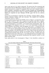
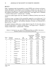
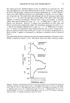
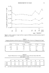
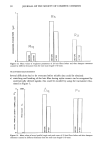
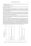
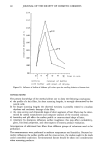
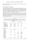
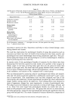
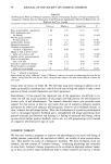
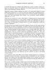
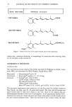
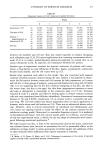
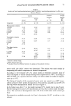
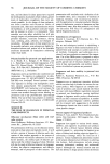
CYTOLOGY OF STRATUM CORNEUM 61 Table III Corneocyte Counts in Trial I (mean and standard deviation) Weeks 0 1 2 3 4 6 Isotretinoin 1.5% m 53,981 81,429 56,344 80,5 12 sd 15,017 43,106 14,138 17,664 Tretinoin 0.05% m 34,733 48,336 72,987 74,820 sd 15,951 22,737 18,382 25,639 Ethanol + m 45,104 45,490 26,291 45,104 propyleneglycol aa sd 29,087 26,430 8,514 22,156 Motretinid 0.5% m 39,557 62,278 60,445 64,931 sd 18,205 29,639 27,035 13,335 Gel base m 34,009 39,074 33,382 24,313 sd 15,506 23,358 15,550 13,368 83 455 1 845 76.521 14 617 33.648 12.192 74 241 8.890 30 488 15,799 46,4O2 15,799 36,569 14,465 38,716 16,974 45,156 29,427 65,900 34,099 between cell numbers and cell size: Skin was treated topically to enhance (stripping with cellophane tape (2,24,25)) topical application of an irritating concentration of reti- noids (2,3) or to reduce epidermopoiesis (glucocorticosteroids on normal skin or on contact dermatitis (2,9)). As expected, the corneocytes followed this pattern. Another type of experiment involved the systemic treatment of patients with isotre- tinoin, a drug known to cause exfoliation of the skin. Again, as postulated, corneocytes became much smaller, and the cell counts went up (12). Several other questions were asked in this model. One was concerned with seasonal variation of human stratum corneum during the year. Indeed, it was possible to charac- terize the fluctuation between warm and cold seasons for both parameters, cell counts and cell size, in four groups of subjects, young and old females and young and old males (10). It is not surprising that the dry skin condition (chapping or dry skin, dry skin in the winter time, dry skin in the aged, dry skin from inappropriate exposure to water and soap or detergents) is measurable in the corneocyte assay (15,31,32). Tretinoin (vitamin-A acid) is a popular and effective peeling agent to remove stratum corneum cells and even comedones from follicular infundibula. Commercial concentrations range between 0.1% and 0.025%. Isotretinoin (13-cis retinoic acid) is a powerful oral anti- acne drug. We (33) have looked into sebum suppression after its topical application in humans, while others used the hairless rat (34). There was no substantial sebum reduc- tion. However, there is clinical improvement of comedonal and inflammatory acne (33), hence our ongoing interest to study the effects on the stratum corneum by the exfoliative cytology model. Motretinid is a somewhat forgotten retinoid. It is marketed in Switzerland and France. Very few clinical studies attest to its efficacy, and we know of only one experimental study, in which the size and shape of corneocytes were investi- gated following motretinid application.* We wish to point out that, at least in the concentrations used in this study, motretinid was tolerated better than tretinoin or isotretinoin but led to measurable effects in the exfoliative cytology model. It seems appropriate to further explore the usefulness of this or other retinoids where exfoliation from topical treatment is intended. * R. Marks, A.D. Pearse, D. Black, and S. Hill, Techniques for assessment of the activity of topically applied retinoids (1986, unpublished personal communication).

62 JOURNAL OF THE SOCIETY OF COSMETIC CHEMISTS REFERENCES (19) (20) (21) (22) (23) (24) (25) (26) (1) K. J. McGinley, R. R. Marpies, and G. Piewig, A method for visualizing and quantitating the desquamation portion of the human stratum corneum, J. Invest. Dermatol., 53, 107-111 (1969). (2) E. H/51zle and G. Plewig, Effect of dermatitis, stripping and steroids on the morphology of corneo- cytes. A new bioassay, J. Invest. Dermatol., 68, 350-356 (1977). (3) S. Lee, Y. K. Park, Y. K. Kim, and J. S. Kang, An experimental study on corneocytes of acutely and chronically irritated skin, Arch. Dermatol. Res., 275, 49-52 (1983). (4) H. Goldschmidt, Surface area measurements of psoriatic corneocytes: Effects of intralesional steroid therapy, J. Invest. Dermatol., 73, 558-560 (1979). (5) M. T. Hojyo-Tomoka and A.M. Kligman, Does cellophane stripping remove the horny layer? Letters to the editor, Arch. Dermatol., 106, 767-768 (1972). (6) R. Hunter, H. Pinkus, and C. H. Steele, Examination of the epidermis by the strip method. III. The number of keratin cells in the human epidermis, J. Invest. Dermatol., 27, 31-34 (1956). (7) E. H/51zle, J. Park, and G. Plewig, EinfluB verschiedener Glukokortikoide und ihrer Grundlagen auf die Korneozyten der normalen Epidermis, Akt. Dermatol., 6, 75-81 (1980). (8) M. Papini, P. Mariani, and P. Calandra, Topical motretinid treatment of skin diseases with patho- logical keratinization, Ann. It. Derm. Clin. Sper., 37, 175-180 (1983). (9) E H/51zle, G. Plewig, and A. Ledolter, "Corneocyte Exfoliative Cytology: A Model to Study Normal and Diseased Stratum Corneum," in Skin Models, 1st ed., R. Marks, and G. Plewig, Eds. (Springer Verlag, Berlin, Heidelberg, 1986), pp. 183-193. (10) S. Herrmann, E. Scheuber, and G. Plewig, "Exfoliative Cytology: Effects of the Seasons," in Stratum Corneum, 1st ed., R. Marks and G. Plewig, Eds. (Springer Verlag, Berlin, Heidelberg, 1983), pp. 181-185. (11) H. Goldschmidt and A.M. Kligman, Exfoliative cytology of human horny layer, Arch. Dermatol., 96, 572-576 (1967). (12) W. Breiner, E. $cheuber, and G. Piewig, "Effects of Isotretinoin (13-cis Retinoic Acid, Ro 4-3780) Treatment on Exfoliative Cytology," in Stratum Corneum, 1st ed., R. Marks and G. Piewig, Eds. (Springer Verlag, Berlin, Heidelberg, 1983), pp. 222-226. (13) G. Piewig, Regional differences of cell sizes in the human stratum corneum. Part II: Effects of sex and age, J. Invest. Dermatol., 54, 19-23 (1970). (14) G. Piewig and R. R. Marpies, Regional differences of cell size in the human stratum corneum, J. Invest Dermatol., 54, 13-18 (1970). (15) R. Marks, Epidermal and corneocyte size changes with age, Brit. Jo Dem., 102, 738-739 (1980). (16) H. Germann, W. Barran, and G. Piewig, Morphology of corneocytes from human nail plates, Jo Invest. Dermatol., 74, 115-118 (1980). (17) K. A. Holbrook and G. F. Odland, Regional differences in the thickness (cell layers) of the human stratum corneum: An ultrastructural analysis, J. Invest. Dermatol., 62, 415-422 (1974). (18) G. Piewig, E. Scheuber, B. Reuter, and W. Waidelich, "Thickness of Corneocytes," in Stratum Corneum, 1st ed., R. Marks and G. Piewig, Eds., (Springer Verlag, Berlin, Heidelberg, 1983), pp. 171-173. F. Allegra and C. De Panfilis, An in vivo method of studying the kinetics of cell proliferation in normal human epidermis, Acta Dermatovener (Stockholm), 54, 87-90 (1974). H. Baker and A.M. Kligman, Technique for estimating turnover time of human stratum corneum, Arch. Dermatol., 95, 408-411 (1967). P. Corcuff, G. Delesalle, and H. Schaefer, Quantitative aspect of corneocytes,J. Soc. Cosmet. Chem., 33, 1-7 (1982). W. L. Epstein and H. I. Maibach, Cell renewal in human epidermis, Arch. Dermatol., 92, 462-468 (1965). H. Goldschmidt and A.M. Kligman, Desquamation of the human horny layer, Arch. Dermatol., 95, 583- 586 (1967). H. Pinkus, Examination of the epidermis by the strip method of removing horny layers. Observation of thickness of the horny layer and on mitotic activity after stripping. J. Invest. Dermatol., 16, 383-386 (1951). H. Pinkus, Examination of the epidermis by the strip method: Biometric data on regeneration of the human epidermis, J. Invest Dermatol., 19, 431-447 (1952). G. Piewig and A. M. Kligman, Zellkinetische Untersuchungen bei Kopfschuppenerkrankung (Pity- riasis simplex capillitii), Arch. Klin. Exp. Derre., 236, 406-421 (1970).
Purchased for the exclusive use of nofirst nolast (unknown)
From: SCC Media Library & Resource Center (library.scconline.org)