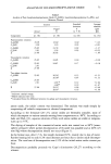
26 JOURNAL OF THE SOCIETY OF COSMETIC CHEMISTS (45) (46) (47) (48) (49) (50) Function," in Stratum Corneum, R. Marks and G. Plewig, Eds. (Springer Verlag, Berlin, Heidelberg, New York, 1983), pp. 175-180. J. L. Lev&que, P. Cotcuff, J. De Rigal, and P. Agache, In vivo studies of the evolution of physical properties of the human skin with age, Int. J. Dermatol., 23(5), 322-329 (1984). H. I. Blank, Factors which influence the water content of the stratum corneum, J. Invest Dermatol., 18, 433-437 (1952). H. I. Blank, J. Moloney, A. G. Emslie, I. Simon, and C. Apt, The diffusion of water across the stratum corneum as a function of its water content, J. Invest Dermatol., 82, 188-194 (1984). W. Montagna and K. Carlisle, Structural changes in aging human skin, J. Invest Dermatol., 73, 47-53 (1979). G. K'tistala, Dermal-epidermal separation: Influence of age, sex and body region on suction blister formation in human skin, Ann. Clin. Res., 4, 10-22 (1972). G. Sauermann and U. Hoppe, The determination of the volume of human epithelium cells. Confer- ence on the scientific basis of skin care. Congress of the International Federation of the Societies of Cosmetic Chemists, Denmark, 1981.

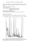
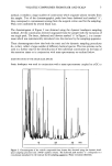
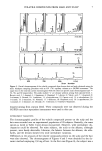
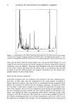
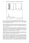
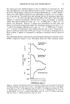
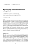
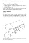
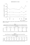
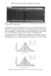
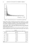
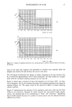
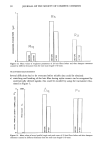
J. Soc. Cosmet. Chem., 39, 27-42 (January/February 1988) Measurement of the surface profile of human hair by surface profilometry G. SAUERMANN, U. HOPPE, R. LUNDERST)•DT, and B. SCHUBERT, Beiersdorf AG, Unnastr. 48, 2000 Hamburg 20, West Germany (G.S., U.H., B.S.), and Universitiit der Bundeswehr Hamburg, Holstenhofweg 85, 2000 Hamburg 70, West Germany (R.L. ). Received Novermber 25, 1986. Synopsis The surface of single hair fibers of Europeans was investigated by profilometry using pyramidal and axe- shaped styli. Measuring parallel to the hair axes from root towards tip, the mainly used scan-lengths were 0.4 mm, 4 mm and 40 mm, yielding roughness-and-waviness data (R a, R v, Rq, R z, Aq). The structure of the recorded profiles (0.4 mm scan length) is determined mainly by the sequence of cuticle cells: the counted peaks (Pc) correspond well with the density of cells/cm hair length. When scanning longer dis- tances, the recorded profile seems to be determined by stochastic events and by the influence of bio- rhythms. Increasing damage at the hair tips could be detected. The reproducibility of the method is excellent. Wet hair shows considerably higher roughness parameters. Influences of shampoos using in vitro and in vivo techniques were investigated. The method to perform transversal measurements which yield information concerning cross-sectional pa- rameters of the hair fibers will be discussed, including possible pitfalls. Buffers of pH 6 increase the diameter of the fiber considerably less than buffers of pH 9. The ellipsoidal parameters of hair fibers can be determined by computer-supported optimal fitting of ellipses into the experimentally recorded contours. Twisting the hair fiber around its main axes causes a helical shape of the fiber the differences between the ellipsoidal axes may possibly be estimated from the longitudinal profile. INTRODUCTION The surface structure of human hair is well known from scanning electron microscopic photos. The dominating structural elements are the orderly arranged cuticle cells which protect the interior of the hair fiber against chemicals, microorganisms, and physical insult. Quantitative interpretation of the hair surface by microscopy is afflicted with especially one disadvantage: dimensions perpendicular to the fiber axis can be deter- mined only by time-consuming tedious work. Furthermore, small quantitative alter- ations--periodic or stochastic and extended over long distances--will presumably not be discovered. Cross-sectional parameters can be determined by optical microscopy the disadvantages are numerous, however, but the advantages lie in the visual recognition of the object to be measured (1). 27
Purchased for the exclusive use of nofirst nolast (unknown)
From: SCC Media Library & Resource Center (library.scconline.org)