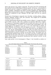
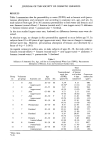
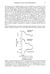
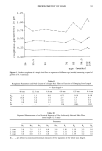
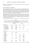
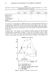
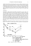
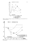
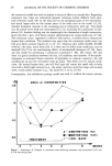
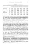
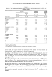
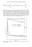
CORNEOCYTE SIZE AND PERMEABILITY 23 of the intercellular spaces due to an increase in the size of the corneal cells. This would be too simplistic an approach, and we should take into account other factors which affect the physical and physicochemical properties of the barrier, such as modifications in the lipid content and/or the lipid composition of the intercellular cement, cohesion between corneocytes (44,45), and the hydration level of the horny layer (46,47). More- over, morphological and functional changes in adjacent structures, in particular the dermis, should also be taken into consideration. Thus, in advancing age, alterations in the vascular bed and extracellular matrix may lead to a decreased clearance of transder- mally absorbed materials from the dermis (48,49). Influence of sex. As our results show (Table I, Figure 2), in the areas studied (upper-outer arm, forehead), no differences were found between male and female subjects, either in percutaneous absorption of benzoic acid or in TEWL. There has been no systematic study showing the effects of sex on cutaneous permeability in man. We, therefore, cannot compare our results with the literature. Some authors (7) report that corneocytes are smaller in men than in women. Others state the contrary (50). We agree with Marks et al. (11) that there do not appear to be any obvious differences in the surface areas of the corneocytes between men and women. TRANSEPIDERMAL WATER LOSS-PERCUTANEOUS ABSORPTION RELATIONSHIP Although most authors recognize the importance of anatomic site either in the degree of absorption or in TEWL, the literature does not include any quantitative data on the relationship which may exist between these two parameters in man. Our results show (Figure 4) that for the anatomic sites studied and within the range of TEWL and penetration values determined, there exists a highly significant linear relationship (r = 0.92, p 0.001) between the permeability of the skin to water and the percutaneous absorption of a non-water-soluble compound such as benzoic acid. Only values obtained on aged subjects (65-80 years, upper-outer arm, no. 3) do not fit this correlation. Thus, although the barrier function of the stratum corneum to the penetration of envi- ronmental agents appears to decline with age, we agree with others that TEWL does not vary (38,44,45). This is a strange situation because a particular feature of aged skin is the roughness and apparent dryness of its surface. As shown in this paper, generally less penetration occurs in those regions with large corneocytes. The larger the corneo- cytes, the smaller the intercellular space. In the case of the elderly, less lipid in the stratum corneum due to a decrease in the volume of the intercellular space could ac- count for the decreased permeation of lipid-soluble compounds like benzoic acid. This explanation is at the same time consistent with the unchanged permeation of water. CONCLUSION In the present work we have demonstrated that in young subjects the two principal indicators of the functional state and integrity of the cutaneous barrier, i.e., percuta- neous absorption and TEWL, are directly linked. Although corneocyte surface area appears to be an important factor in the efficiency of the epidermal barrier in both these phenomena, our results show that it only partly explains the differences in permeability observed according to anatomic site or age. It seems that other factors are relevant when corneocyte surface area is less than 600 p,m 2 or more than 1000 p,m 2. Thus, worthy of

24 JOURNAL OF THE SOCIETY OF COSMETIC CHEMISTS research, and being studied at the moment, are changes in the lipid content of the intercellular spaces which could affect the whole physicochemistry of the cutaneous barrier. ACKNOWLEDGEMENTS The authors would like to thank A. M. Cabaillot and J. MacMaster for their excellent technical assistance. REFERENCES (1) D. R. Wilson and H. I. Maibach, "A Review of Transepidermal Water Loss: Physical Aspects and Measurements as Related to Infants and Adults," in Neonatal Skin, H. I. Maibach and E. K. Boisits, Eds. (Marcel Dekker, New York, 1982), pp. 83-100. (2) H. I. Maibach, R. Bronaugh, R. Guy, E. Turr, D. R. Wilson, S. Jacques, and D. Chaing, "Non- Invasive Techniques for Determining Skin Function," in Cutaneous Toxicity, V. A. Drill and P. Lazar, Eds. (Raven Press, New York, 1984), pp. 63-97. (3) D. Dupuis, A. Rougier, C. Lotte, D. R. Wilson, and H. I. Maibach, In vivo relationship between percutaneous absorption and transepidermal water loss according to anatomic site in man, J. Soc. Cosmet. Chem,, 37, 351-357 (1986). (4) C. Lotte, A. Rougier, D. R. Wilson, and H. I. Maibach, In vivo relationship between transepi- dermal water loss and percutaneous penetration of some organic compounds in man: Effect of ana- tomic site, Arch Dermatol. Res. (in press). (5) }-I. I. Maibach, R. J. Feldmann, T. Milby, and W. Serat, Regional variations in percutaneous pene- tration in man, Arch. Environ. Health, 23, 208-211 (1971). (6) A. Rougier, C. Lotte, and }-I. I. Maibach, In vivo percutaneous penetration of some organic com- pounds related to anatomic site in man: Predictive assessment by the stripping method, J. Pharm. Sci., 76, 451-454 (1987). (7) G. Piewig and B. M. Marples, Regional differences of cell sizes in the human stratum corneum. Part I,J. Invest. Dermatol., 54, 13-18 (1970). (8) G. Plewig, Regional differences of cell sizes in the human stratum corneum. Part II: Effect of sex and age, J, Invest. Dermatol., 54, 19-23 (1970). (9) G. L. Grove, Exfoliative cytological procedures as a non-intrusive method for dermatogerontological studies,J. Invest. Dermatol., 79, 67-69 (1979). (10) G. L. Grove, R. M. Lavker, E. Holzle, and A.M. Kligman The use of non-intrusive tests to monitor age associated changes in human skin, J. Soc. Cosmet. Chem., 32, 15-26 (1981). (11) R. Marks, S. Nicholls, and C. S. King, Studies on isolated corneocytes, Int. J. Cosm. Sci., 3, 251-258 (1981). (12) R. L. Bronaugh, R. F. Stewart, E. R. Contigon, and A. L. Giles, Methods for in vitro percutaneous absorption studies. I: Comparison with in vivo results, Toxicol. Appl, Pharmacol., 62, 474-480 (1982). (13) J. W. Bridges, M. R. French, R. L. Smith, and R. T. Williams, The fate ofbenzoic acid in various species, Biochem. J., 118, 47-51 (1970). (14) R. J. Feldmann and H. I. Maibach, Absorption of some organic compounds through the skin in man, J. Invest. Dermatol., 54, 339-404 (1970). (15) A. Rougier, D. Dupuis, C. Lotte, R. Roguet, and }-I. Schaefer, In vivo correlation between stratum corneum reservoir function and percutaneous absorption, J. Invest. Dermatol., 81, 275-278 (1983). (16) D. Dupuis, A. Rougier, R. Roguet, C. Lotte, and G. Kalopissis, In vivo relationship between horny layer reservoir effect and percutaneous absorption in human and rat, J. Invest. Dermatol., 82, 353-356 (1984). (17) K.J. McGinley, R. R. Marples, and G. Plewig, A method for visualizing and quantitating the desquamating portion of the human stratum corneum, J, Invest. Dermatol., 53, 107 - 111 (1969). (18) P. Corcuff, G. Delesalle, and H. Schaefer, Quantitative aspect of corneocytes. J. Soc. Cosmet. Chem., 33, 1-7 (1982).
Purchased for the exclusive use of nofirst nolast (unknown)
From: SCC Media Library & Resource Center (library.scconline.org)