276 JOURNAL OF THE SOCIETY OF COSMETIC CHEMISTS products and that testing will be required prior to commercialization. It would be particularly appropriate to have an animal model for prescreening with UVA sunscreens because to observe the biologic effects in the human requires either an extended expo- sure period or a presensitization with a phototoxic/photocarcinogenic psoralen (8,9). The guinea pig has been shown to yield well-defined, reproducible erythema to ultravi- olet B irradiation (UVB, 290- 320 nm) (10,11) and to ultraviolet A irradiation (UVA, 320-400 nm) (12), especially when predosed with 8-methoxypsoralen (8MOP) (3). The other animal model most suggested is the mouse, but this model does not develop marked erythema, and the investigator must rely on the development of edema as an endpoint evaluation. This is perhaps due to the fact that the guinea pig mast cell is a histamine releaser, whereas the mouse mast cell predominantly releases serotonin (1) the latter is known to produce edema in the rodent. Additionally, the guinea pig has a broad surface area allowing for multiple-site testing and its hair can provide an effective "blocking" template on areas where exposure is unwanted. It has also been shown to exhibit a high degree of correlation to the human response when used under both static and wash-off conditions (10, 11, 14). For these reasons, we selected it for use in preclin- ical sun protection studies. With regard to testing sunscreens for UVA efficacy, there is considerable controversy with regard to the proper approach (8,16). One of the commonly used approaches is to use the phototoxin/photocarcinogen 8MOP as a means to increase the human subjects' susceptibility to long-wave ultraviolet radiation. This should be considered an unaccep- table risk for these volunteers, except for very unusual circumstances. The other tech- niques (e.g., immediate tanning, delayed tanning) require extensive irradiation times and are expensive to run. Psoralen, dosed orally, results in a severe phototoxic reaction when animals or humans are exposed to the shorter wavelengths of UVA (320-340 nm, peak @ 335 nm). Although there is some question (8) as to whether testing at the shorter wavelengths of UVA (17) is proper for purposes of determining an SPF, it seems logical and appropriate to use this approach, since the shorter wavelengths are the most biologically active. Therefore, a methodology has been developed as a screening proce- dure that will identify the potential of a material to protect against UVB or UVA radiation in accordance with its expected clinical "sun protection factor." Since this clinical "sun protection factor" most often represents the protection value analyzed, we have defined this factor to be the "target value" for use in our assays. It must be stressed that label claims on sunscreen products are based solely on the results of human testing (14). It is not our suggestion that this screening procedure replace the human assay, but that it serve as an aid to identify raw materials and formulations worthy of subsequent evaluation at the clinical level. MATERIALS AND METHODS SUNSCREEN PREPARATIONS Eight experimental or proprietary sunscreen formulations were submitted to our labora- tory for testing. The data were to be compared to sun protection factors (SPF) derived from clinical evaluations conducted according to the FDA monograph (14). In addition, 8% homosalate, recognized by the FDA as a standard for clinical trials, was included in this investigation. Some of these materials were additionally tested in human panels by

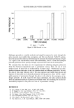
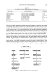
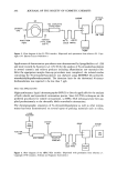
SUNSCREEN EFFICACY 277 Hill Top Research Inc. for confirmatory clinical data, according to the FDA monograph (14). ANIMAL PREPARATION Two young adult male Hartley guinea pigs from Murphy Breeding Laboratories Inc. (Plainfield, IN), weighing approximately 750 grams each, were used for each trial. The large size of this animal provided a broad dorsal surface area for exposure and irradia- tion, although this size of animal is not critical to the observation. On the day prior to treatment, the entire dorsal surface of each animal was clipped using a no. 40 clipper blade. This area was then depilated using a commercial depila- tory that was applied and left for no more than 15 minutes. The depilatory was washed off thoroughly with warm running water. The animals were towel dried and returned to their cages. TEST MATERIAL APPLICATION On the day following animal preparation, a rectangular area (approximately 3 X 10 cm), parallel to the dorsal midline on the right side of each animal, was delineated with a waterproof marker. Using a microliter syringe, the test material was deposited evenly across the rectangle, resulting in a total application of 2 I•l/cm 2. This material was spread using a gloved fingertip. For the evaluation of UVA screening potential, approx- imately two hours prior to test material treatment, 8MOP was administered orally at a dose of 20 mg/kg. This rendered the animals photosensitive to the UVA spectrum. IRRADIATION The animals were restrained on an irradiation table in a circular arrangement (15). After the animals had been properly positioned, opaque templates were applied, isolating three test sites, arranged linearly, on each side of the dorsal midline. A fourth skin site was isolated on one side of each animal as a nonirradiated primary irritation control site. A light wheel (15), holding either twelve UVA black light bulbs (20 watt F20T12/BL) or twenty four UVB sunlamp bulbs (FS20 USA, 20 watt) in a spoke-like fashion, was started and revolved over the restrained animals. Approximately two minutes after the wheel had been started, the lamps were turned on. The three untreated test sites on the left side of the animal were sequentially irradiated from head to tail for three, six, and nine minutes, respectively, when testing with UVB radiation and for six, eight, and ten minutes, respectively, when testing with UVA radiation. The exposed skin sites were occluded, as appropriate, with plastic tape to discontinue exposure. The middle time value had been predetermined to approximate the respective minimal erythemic dose (MED) for the experimental conditions. The opposite three treated test sites were each irradiated for time periods that corresponded to the product of the target value to be investigated and the irradiated time period of the paired untreated site. WASH-OFF When wash-off data were to be obtained, the appropriate animals were individually
Purchased for the exclusive use of nofirst nolast (unknown)
From: SCC Media Library & Resource Center (library.scconline.org)