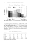
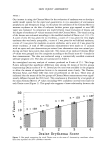
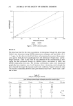
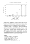
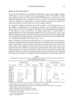
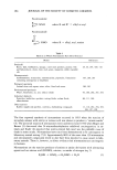
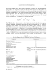
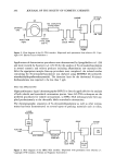
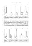
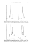
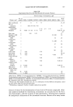
264 JOURNAL OF THE SOCIETY OF COSMETIC CHEMISTS • 15 • lO z• o v/-/• 4 Min • õ Min 0.5 1 1.5 2 3 4 5 6 24 TIME in Hours Figure 3. Mean a* chromaticity coordinate values at each observation time for 4- and 8-minute mustard vapor exposures on hairless guinea pigs. N = 33 error bars = _+ 95% confidence intervals. Draize scores and the measured Chroma Meter values. The Spearman correlation coeffi- cient was calculated to be 1.00, and it is statistically significant. The time course of developing erythema as measured by the mean Chroma Meter a* values for the 4- and 8-minute HD vapor exposures is illustrated in Figure 3. The 95% confidence intervals and the corresponding Draize scores are also included on the bar graph. These data indicate that erythema was first observed at about 2 hours and reached a maximum between 4 and 6 hours. By 24 hours there was a slight decrease. At the 6-hour observation time the mean a* Chroma Meter values of the 4- and 8-minute HD exposures were 15.0 and 16.7, while the median Draize scores were 3 and 4, respectively. This represented a significant difference at the 95% level. Other analytical methods of lesion evaluation, including high-resolution ultrasound, laser Doppler flow measurements, and transepidermal water loss, would be useful but are not yet available in our laboratory. We plan to acquire and evaluate these methods in the future. CONCLUSIONS We have demonstrated that the Minolta CR-200 Chroma Meter is capable of providing a reproducible and quantitative assessment of the degree of erythema produced on the skin of euthymic hairless guinea pigs exposed to HD vapor. Furthermore, the a* chro- maticity coordinate very closely parallels the visual Draize scores. We have demon- strated for the first time that an objective analytical method can be used for the assess- ment of skin damage caused by HD exposure. We plan to develop this technique into a validated in vivo model for the assessment of antivesicant prophylactic and therapeutic drugs.

SKIN INJURY ASSESSMENT 265 REFERENCES (1) J. W. Feather, K. S. Ryarr, J. B. Dawson, J. A. Cotterill, D. J. Barker, and D. J. Ellis, Reflec- tance spectrophotometric quantificarion of skin colour changes induced by topical corticosteroid prep- arations, Br. J. DermatoL, 106, 437 (1982). (2) B. L. Diffey, R. J. Oliver, and P.M. Farr, A portable instrument for quantifying erythema induced by ultraviolet radiation, Br. •. DermatoL, 111, 663 (1984). (3) S. W. Babulak, L. D. Rhein, D. D. Scala, F. A. Simion, and G. L. Grove, Quantirarion of ery- rhema in a soap chamber test using the Minolta Chroma (Reflectance) Meter: Comparison of instru- mental results with visual assessments, J. $oc. Cosmet. Chem., 37, 475 (1986). (4) P. Bierring and P. H. Andersen, Skin reflectance spectrophotometry, Photodermatology, 4, 167 (1987). (5) T. Nose, A. Uchiyama, and K. Tsurumi, Study on the ultraviolet eryrhema in guinea-pigs, Jpn. J. PharmacoL, 46, 296 (1988). (6) J. C. $eitz and C. G. Whirmore, Measurement of erythema and tanning responses in human skin using a tristimulus colorimeter, Dermatologica, 177, 70 (1988). (7) W. Westerhof, O. Estevez-Uscanga, A. Kammeyer, J. Meens, M. Durocq, and I. Cair, Quantitative skin reflectance chromameter measurements of erythema and pigmentation response in human volun- teers of different skin types irradiated with solar simulated UV, J. Invest. Dermato/., 91, 385 (1989). (8) K. P. Wilhelm, C. Surber, and H. I. Maibach, Quantification of sodium lauryl sulfate irritant der- matitis in man: Comparison of four techniques: skin color reflectance, transepidermal water loss, laser Doppler flow measurement and visual scores, Arch. Dermatol. Res., 281, 293 (1989). (9) W. Westerhof, O. Estevez-Uscanga, A. Candido, H. Coevoet, and A. Kammeyer, Separation of erythema and pigmentation measurements by means of a skin reflectance chromameter, C/in. Res., 37, 736A (1989). (10) W. Westerhof, O. Estevez-Uscanga, A. Candido, H. Coevoet, and A. Kammeyer, Separation of erythema and pigmentation measurements by means of a skin reflectance chromameter, J. Invest. Dermatol., 92, 540 (1989). (11) K. P. Wilhelm and H. I. Maibach, Skin color reflectance measurements for objective quantification of erythema in human beings, J. Am. Academy Dermatol., 21, 1306 (1989). (12) E. Berardesca and H. I. Maibach, Alternative nonvisual methods quantitating skin test response, Immun. Allergy Clinics North Am., 9, 597 (1988). (13) Minolta Corporation, 101 Williams Dr., Ramsey, New Jersey 07446. (14) D. D. Marlow, M. M. Mershon, L. W. Mitcheltree, G. P. Jaax, and J. P. Petrali, Sulfur mustard induced skin injury in hairless guinea pigs, J. Toxicol., Cutaneous Ocul. Toxicol., 9(3), (1990). (15) G. Wyszecki and W. S. Stilles, Color Science: Concepts and Methods, Quantitative Data and Formulas, 2nd ed. (John Wiley and Sons, New York, 1982) pp. 165-168. 16) R. W. G. Hunt, Measuring Colour (Ellis Horwood Limited, Chichester, England, 1987), pp. 32-130. 17) Vetalar TM, 100 mg/ml. Park Davis, Division of Warner-Lambert Co., Morris Plains, NJ 07950. 18) Rompan, 20 mg/ml. Mobay Corp., Animal Health Division, Shawnee, KS 66201. 19) J. H. Draize, G. Woodard, and H. O. Calvery, J. Pharmacol. Exp. Ther., 83, 377 (1944). (20) J. A. Draize, Appraisal of the Safety of Chemicals in Foods, Drugs, and Cosmetics--Dermal Toxicity (Assoc of Food and Drug Officials of the US, Topeka, Kansas, 1965), pp. 49-52. (21) Method of Testing Primary Irritant Substances (16 CRF, Ch. II, Part 1500.41., 1 Jan 1987), pp. 348-349. (22) J. L. Hintze, Number Cruncher Statistical System (Dr. Jerry L. Hintze, Kaysville, Utah, 1987), pp. 167-190.
Purchased for the exclusive use of nofirst nolast (unknown)
From: SCC Media Library & Resource Center (library.scconline.org)