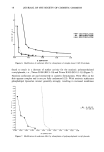
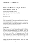
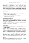
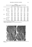
72 JOURNAL OF THE SOCIETY OF COSMETIC CHEMISTS Other membrane components, such as proteins, are embedded in it and depend on the fluidity of the lipid membrane for carrying out many of their functions. For this reason liposomes have been widely used as model membrane systems since their original conception (6,7) to study factors affecting membrane transport, membrane integrity, and disruption. In assessing the damaging effects of extraneous agents on cells, the use of liposomes greatly simplifies dealing with the complexity and variable composition of in vivo systems, while the basic properties of lamellar lipid bilayers common to all cellular membranes are retained. This report describes the development of an in vitro indicator of surfactant irritation on the skin, measuring leakage of a fluorescent marker from phospholipid liposomes ex- posed to surfactant solutions. This method is objective and sensitive and is shown to be capable of indicating the relative irritation potential of a series of anionic surfactants and anionic blends. A mathematical index is developed to facilitate correlation of the data with currently used in vivo indices. It is not expected that the liposome method alone would replace any of the in vivo methods currently in use it may, however, serve as one of a battery of tests that together would give a comprehensive assessment of skin irritancy. The use of liposomes for this particular application is further indicated by findings that correlate skin irritancy directly with the ability of a surfactant system to solubilize skin lipids (8,9). Removal of skin lipids, in turn, leads to a variety of undesirable symptoms, which range from dryness, tightness, scaling, itching, and burning to cracking, in- flammatory changes and, eventually, to a breakdown in the barrier function of the skin (10-12). MATERIALS AND METHODS Egg phosphatidylcholine was purchased from Sigma Chemical Co., St. Louis (Type V-E, 99% in chloroform solution), cholesterol from Nucheck Prep, Elysian, Minnesota, and dicetylphosphate from Aldrich Chemical Co., Milwaukee, Wisconsin. The reference compounds taurodeoxycholate and sodium dodecyl sulfate were purchased from Calbi- ochem, LaJolla, California, and BDH Chemicals Ltd, Poole, U.K., respectively. 5(6)- Carboxy fluorescein (Eastman Kodak) was further purified prior to use (13,14). The surfactants tested were of industrial grade from various sources and were used as sup- plied. PREPARATION OF LIPOSOMES (FIGURE 1) Unilamellar liposomes were produced by the petroleum ether evaporation technique adapted from that described by Schieren et al. (15). Water-jacketed glass chambers resembling micro condensers were custom made by D&H Glassblowing, West Bend, Wisconsin. Phosphatidyl choline, dicetyl phosphate, and cholesterol, in a ratio of 7:2:1 (16,17), were dissolved in chloroform to which a minimal amount of methanol was added. The mixture was then dried under reduced pressure at 45øC for several hours. The dried residue was kept under argon at 4øC until use, when it was dissolved in 35 ml of petroleum ether (Aldrich Chemical Co.) (boiling range: 35-60øC). The solution was then divided evenly into two gas-tight syringes (Hamilton, Reno, Nevada) and

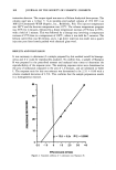
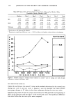
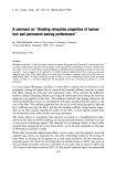
SURF. ACTANT-SKIN INTERACTIONS 73 labtAKE L Figure 1. Liposome preparation by petroleum ether evaporation. injected simultaneously into the two glass chambers that contained 4 ml each of the trapping solution at 60øC. The injection rate was controlled by an injection pump (Sage Instruments) to approximately 0.2 ml/min. Liposomes thus produced were always used within three days. Aliquots for each day's work were separated at this point and refrigerated. TRAPPING SOLUTION-ENCAPSULATION OF FLUORESCENT DYE IN INTERIOR OF INTACT LIPOSOMES (FIGURE 2) The trapping solution was prepared to contain 0.01 M 5(6)-carboxyfluorescein in 0.05 M Tris buffer at pH 7.4. Carboxyfluorescein fluorescence is quenched at higher con- centrations. Thus, leakage, i.e., dilution of the dye, is measured as increase in fluores- cence intensity. pH and temperature also affect the fluorescence readings. It is therefore essential to standardize osmolality (to prevent dilution of the dye caused by water movement into or out of the liposomes), pH, and temperature, so that membrane stability relates directly to the surfactant concentrations in the respective test solutions. Thus, the osmolality of not only the trapping solution, but also of every other solution coming into contact with the liposomes was adjusted to 290 mOsm/kgH20 with NaCI. The osmolality was measured with a Wide Range Osmometer (Advanced Instruments, Inc., Needham Heights, Mass.). Test solutions (4 ml) were pre-heated to 37øC, and a temperature-stabilized sample cell holder in a Perkin Elmer 650S fluores- cence detector was used for the fluorescence determinations. The excitation and emission wavelengths were 492 nm and 520 nm, respectively. Prior to use the external trapping solution was removed by gel chromatography (Seph- adex G-25). 60-Drop aliquots were collected, and the first and last fractions containing liposomes were discarded to assure an even size distribution.
Purchased for the exclusive use of nofirst nolast (unknown)
From: SCC Media Library & Resource Center (library.scconline.org)