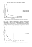
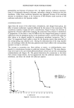
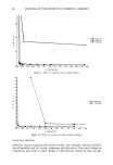
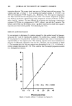
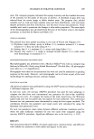
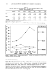
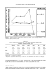
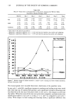
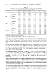
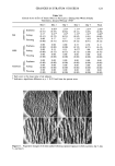
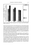
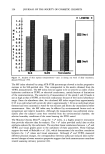
CHANGES IN STRATUM CORNEUM 127 The L* index, which measures brightness of the skin surface, appears to be less useful. The skin brightness may be affected by the degree of erythema of the skin. Also, the L* values of normal skin fall within a fairly narrow range that limits measurement sensi- tivity. Visual assessment of the skin by an experienced grader continues to be a very important method for assessing changes in skin condition. Visual grading in this study differen- tiated the degree of the changes caused by the three test surfactants with respect to erythema, scaling, and fissuring. The visual grading also differentiated the changes caused by seasonal variation within the SLS and SLES subgroups. However, the sub- jective nature of visual grading and the use of a three- or four-point grading scale limits the precision of the data for any detailed analysis. Both skin replica images and macrophotography are useful for documenting the skin surface and provide the possibility for subsequent review of the skin condition. For such documentation, it is important to record the images in a highly standardized manner to minimize any variability introduced through the recording process. Current photo- graphic and video technology make it possible to accurately capture the appearance of the skin, and silicone replicas preserve the textural features for subsequent study. Image analysis further provides useful objective quantitative data. In this study, the mean ratio of the shadow area to the shadow perimeter provided the only significant value. This value was increased after SLS treatment in both test cycles, and indicated increases in the mean depth of the dermatoglyphics. Analysis of other quantitative parameters available through image analysis (such as the total area of shadow and total length of shadow perimeter) yielded no meaningful results. Among the three surfactants tested, SLS caused the most severe damage to the skin and, therefore, had the highest irritancy potential. All the parameters used in this study differentiated the skin sites treated with SLS from the other surfactant-treated and control sites. SLES showed milder damage to the skin than SLS, and its slight irritancy potential was best demonstrated by the results from TEWL and electrical conductance measurements. No significant differences were noted between any sites treated with PEG-20 glyceryl monotallowate and their corresponding water-treated control sites, except for minor changes in TEWL and electrical conductance measurements. PEG-20 glyceryl monotallowate induced no significant adverse effects and, therefore, was rated to have no irritancy potential. The test protocol of repeated patch applications and daily measurements, using the test battery of multiple instrumental methods, morphometric measurements, and clinical observations served well to predict irritancy potential and to adduce some mechanisms of damage to the SC resulting from surfactant treatment. REFERENCES (1) A.M. Kligman and W. M. Wooding, A method for the measurement and evaluation of irritants on human skin, J. Invest. Dermatol., 49, 78-94 (1967). (2) F. R. Bettley, The irritant effect of detergents, Tram. St. Johm HospitalDem. Soc., 58, 65-74 (1972). (3) K. E. Malten, Thoughts on irritant contact dermatitis, Contact Dermatitis, 7, 238-247 (1981). (4) C. G. T. Mathias and H. I. Maibach, Dermatotoxicology monographs. I. Cutaneous irritation: Fac- tors influencing the response to irritants, Clinical Toxicology, 13, 333-346 (1978).

128 JOURNAL OF THE SOCIETY OF COSMETIC CHEMISTS (10) (tl) (12) (13) (14) (15) (16) (5) P. G. M. Van der Valk, J. P. Nater, and E. Bleumink, Skin irritancy of surfactants as assessed by water vapor loss measurements, J. Invest. Dermatol., 82, 291-293 (1984). (6) T. Frodin and C. Anderson, Multiple parameter assessment of skin irritancy, Contact Dermatitis, 17, 92-99 (1987). (7) D. Van Neste, G. Mahmoud, and M. Masmoudi, Experimental induction of rough dermatitic skin in humans, Contact Dermatitis, 16, 27-33 (1987). (8) R. J. Scheuplein and L. Ross, Effects of surfactants and solvents on the permeability of epidermis, J. Soc. Cosmet. Chem., 21, 853-873 (1970). (9) G. Imokawa, S. Akassaki, Y. Minematsu, and M. Kawai, Importance of intercellular lipids in water-retention properties of the stratum corneum: Induction and recovery study of surfactant dry skin, Arch. Dermatol. Res., 281, 45-51 (1989). A. W. Fulmer and G. J. Kramer, Stratum corneum lipid abnormalities in surfactant-induced dry scaly skin, J. Invest. Dermatol., 86, 598-602 (1986). P. J. Frosch and A.M. Kligman, The soap chamber test,J. Am. Acad. Dermatol., 1, 35-41 (1979). E. J. Van Scott and L. B. Lyon, A chemical measure of the effect of soap and detergents on the skin, J. Invest. Dermatol., 21, 199-203 (1963). F. R. Bettley, The influence of detergents and surfactants on epidermal permeability, Br. J. Derma- tol., 77, 98-100 (1965). D. C. F. Wood and F. R. Bettley, The effect of various detergents on human epidermis, Br. J. Dermatol., 84, 320-325 (1971). G. Imokawa, K. Sumura, and M. Katsumi, Study on skin roughness caused by surfactants. I. A new method in vivo for evaluation of skin roughness, J. Am. Oil Chem. Soc., 52, 479-483 (1975). R. A. Tupder, J. Pinnagoda, P. Coenraads, and J. P. Nater, The influence of repeated exposure to surfactants on the human skin as determined by transepidermal water loss and visual scoring, Contact Dermatitis, 20, 108-114 (1989). (17) A. Triebskorn, M. Gloor, and F. Greiner, Comparative investigations on the water content of the stratum corneum using different methods of measurement, Dermatologica, 167, 64-69, 1983. (18) S. Freeman and H. I. Maibach, Study of irritant contact dermatitis produced by repeated patch test with sodium lauryl sulfate and assessed by visual methods, transepidermal water loss, and laser Doppler velocimetry, J. Am. Acad. Dermatol., 19, 496-502 (1988). (19) L. L. Hantman, Methods for studying the skin surface, The 1983 IFSCC/SCC Joint Conference on Skin, San Francisco. (20) G. E. Nilsson, Measurement of water exchange through the skin, Med. Biol. Eng. Comput., 15, 209-218 (1977). (21) J. Pinnagoda, R. A. Tupker, T. Agner, and J. Serup, Guidelines for transepidermal water loss (TEWL) measurement: A report from the standardization group of the European Society of Contact Dermatitis, Contact Dematitis, 22, 164-178 (1990). (22) H. Tagami, M. Ohi, K. Iwatsaki, Y. Kanamaru, M. Yamada, and B. Ichijo, Evaluation of skin surface hydration in vivo by electrical measurements, J. Invest. Dermatol., 75, 500-507 (1980). (23) N. A. Puttnam, Attenuated total reflectance studies of the skin,J. Soc. Cosmet. Chem., 23, 209-226 (1972). (24) S. W. Babulak, L. A. Rhein, D. D. Scala, F. A. Simion, and G. L. Grove, Quantitation oferythema in a soap chamber test using the Minolta Chroma (Reflectance) Meter: Comparison of instrumental results with visual assessments, J. Soc. Cosmet. Chem., 37, 475-479 (1986). (25) SAS User's Guide: Statistics (SAS Institute, Inc., Cary, NC, 1982). (26) S. P. Barton and D. R. Black, "Surface Contour: Variability, Significance and Measurements," in The Physical Nature of the Skin, R. M. Marks, S. P. Barton, and C. Edwards, Eds. (MTP Press, Boston, 1988), pp. 23-30. (27) L. M. Mullen, Evaluation of the Moisturizing Characteristics of Ethoxylated Coconut and Tallow Monoglyc- erides, M. S. Thesis, University of Cincinnati (1987).
Purchased for the exclusive use of nofirst nolast (unknown)
From: SCC Media Library & Resource Center (library.scconline.org)