150 JOURNAL OF THE SOCIETY OF COSMETIC CHEMISTS membranes. Knowing that liposomes can be prepared from the interstitial lipids of the stratum corneum, it appears reasonable to attempt modeling the permeability charac- teristics of these lipids using such structures. In fact, liposomes prepared from a lipid mixture consisting of epidermal ceramides, carnauba fatty acids, palmitic acid, choles- terol, and cholesterol sulfate were previously employed for this purpose by Abraham and Downing (7). They formed a membrane by addition of the liposomal suspension to a Millipore © filter backing. The low water flux through the membrane suggested that it was an effective barrier toward water diffusion according to the methodology used for assessment. However, the system suffers the constraint of requiring epidermal cer- amides, a lipid mixture obtained by a difficult and time-consuming procedure involving extensive extraction and isolation from epidermal lipids. The purpose of this study is to investigate the feasibility of preparing a bilayer mem- brane system for use as a model for the lipoidal phase of the stratum corneum using commercially available (semi)synthetic lipids. By utilizing an appropriate lipid mixture whose composition and interlipid interaction strength is similar to those of stratum corneum lipids, liposomes can be prepared and tailored for the preparation of model membranes. Reconstitution of membranes from these liposomes, which will be referred to as "skin lipid" liposomes, may also allow us to define accessible requirements that are necessary to mimic the barrier properties of the skin. In this paper, the methods and conditions used for the preparation of such liposomes are described. Attention is also focused on the miscibility of different lipid species and how variations in lipid compo- sitions may affect the physical characteristics of the resultant liposomes. These will be characterized in terms of their trapped volume and bilayer-forming ability. The bilayer- bilayer interaction potential of the "skin lipid" liposomes will be assessed by monolayer studies. The results are then compared with those of liposomes prepared from lipids extracted from the stratum corneum in order to obtain an optimized synthetic lipid mixture. MATERIALS AND METHODS MATERIALS Palmitic acid, ceramides type III from bovine brain, cholesterol, cholesterol sulfate, and N-2-hydroxyethylpiperazine-N'-2-ethanesulfonic acid (HEPES)-free acid were pur- chased from Sigma Chemical Company (St. Louis, MO). The lipids were of the highest purity grade available. The radioactive markers, [3HI-cholesterol (specific activity 46 Ixci/mmol) and [•4C]-palmitic acid (specific activity 48 Ixci/mmol), were obtained from Amersham (Arlington Heights, IL). These lipids showed a single spot on thin layer chromatography and were used without further purification. 16-Anthroyl oxyl palmitate and 5,6 carboxyfluorescein were obtained from Molecular Probe (Eugene, OR). They were chromatographically purified. Otherwise, all chemicals and solvents used in the experiments were of analytical grade. METHODS Preparation of liposomes from synthetic lipids Liposomes were prepared from commercially available lipids using two methods:

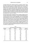
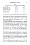
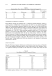
PREPARATION OF LIPOSOMES 151 Conventional hydration method. Appropriate quantities of ceramides, cholesterol, choles- terol sulfate, and palmitic acid were combined using predetermined ratios to obtain lipid mixtures whose compositions are tabulated in Table I. Trace amounts of [•4C]- palmitic acid and [3HI-cholesterol were included as lipid markers. A 1:1 chloroform- methanol mixture was used to dissolve the lipids. A thin film was formed after solvent removal by rotary evaporation (Rotovap ©, Buchi, Switzerland). The trace solvent was removed in vacuo overnight. The lipid film was hydrated at 55øC for 45 min with 0.005 M HEPES buffer, pH 7.50, containing 0.1 M NaC1, 0.001 M EDTA (Fisher Scientific Co., Fairlawn, NJ) and 0.001 mM of the water-soluble marker carboxyfluorescein. After hydration was complete, the liposomes were allowed to anneal at the same temperature for an additional 30 min. The final suspension contained approximately 15 mg/ml lipid. All experiments were carried out in triplicate. Reverse-phase evaporation method. Appropriate amounts of ceramides, cholesterol, palmitic acid, and cholesterol sulfate were dissolved in a l-liter round-bottom flask using a 1:1 chloroform-methanol mixture. Trace amounts of palmitic acid and cholesterol markers were also dissolved in the solvent mixture. HEPES buffer, pH 7.50, containing 0.001 mM carboxyfluorescein was then added to the organic solution. The ratio of the organic solution to the buffer solution was 2:1 (v/v). The mixture, which appeared opalescent, became clear after 18 min sonication (Branson Sonicator E-Module, Shelton, CT). The organic solvent was then removed on a rotary evaporator at 55øC. It was found that the reproducibility of the quantities of lipids incorporated into liposomes was within 3% when the rate of solvent removal was controlled by appropriate adjustment of the vacuum system. After solvent removal was complete, usually within 45-50 min at 55øC, the volume of suspension was adjusted with prewarmed distilled water so that the final suspension contained 30 mg/ml lipid. The liposomes were allowed to anneal in the 55øC bath for an additional 30 min. Table I Lipid Compositions Used to Prepare Liposomes Formulation Ceramides Cholesterol Palmitic Cholesterol number (wt %) (wt %) acid (wt %) sulfate (wt %) 1 46.1 28.9 25.0 0.0 2 40.0 25.0 25.0 10.0 3 30.8 19.2 25.0 25.0 4 15.4 9.62 25.0 50.0 5 0.0 0.0 25.0 75.0 6 43.1 26.9 5.00 25.0 7 40.0 25.0 10.0 25.0 8 33.9 21.1 20.0 25.0 9 15.4 9.6 50.0 25.0 10 0.0 0.0 75.0 25.0 11 46.1 28.9 0.0 25.0 12 0.0 0.0 100.0 0.0 13 49.2 30.8 20.0 0.0 14 55.4 34.6 10.0 0.0 15 0.0 0.0 90.0 10.0 16 0.0 0.0 10.0 90.0
Purchased for the exclusive use of nofirst nolast (unknown)
From: SCC Media Library & Resource Center (library.scconline.org)