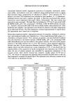
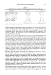
152 JOURNAL OF THE SOCIETY OF COSMETIC CHEMISTS Preparation of liposomes from lipids extracted from stratum corneum of mouse skin Liposomes were prepared from lipids extracted from the stratum corneum as follows. Eight male hairless mice, each sixty days old, were sacrificed in a carbon dioxide chamber. Pieces of full-thickness skin were obtained from both dorsal and abdominal sites. Subcutaneous fat was removed, and the epidermal tissue was separated by im- mersing the entire skin sheet in a Zip-Loc © bag in a 60øC-water bath for 1 min. The tissue was incubated for 2 hr at 37øC on a filter paper saturated with 1% trypsin (Sigma Chemical Co., St. Louis, MO) in phosphate-buffered saline (PBS) containing 30 mM Na2HPO 4, 35 mM NaCI, pH 7.4. The mixture was then washed extensively with mild vortexing to remove granular cells. The stratum corneum was then collected and used for extraction. Lipids were extracted from several stratum corneurn sheets using a modified method developed by Bligh and Dyer (8) and Elias et al. (9). The sample was minced with scissors and extracted overnight in a mixture of chloroform-methanol-water (1:1:0.8 v/v) on an orbit shaker (Labline Instrument Inc., Melrose Park, IL) vibrated at 1,000 rpm. The mixture was then filtered, and the tiltrate was converted to two phases by the addition of equal volumes of chloroform and 0.1 M KCI. The two-phase system was separated by centrifugation at 2,000 rpm for 10 min. The lower phase was collected and washed twice with chloroform:methanol:0.1 M KCI (1:1:0.9 v/v). The upper phases ere combined and sequentially extracted using 1:1 and 2:1 chloroform:methanol solution. The organic phase was collected and combined with the previously washed lower phase. The solvent was then removed by rotary evaporation. Trace amounts of radioactive markers [ 4C]-palmitic acid and [3H]-cholesterol were added to the 15-mg portion of the above-extracted lipid. Liposomes were prepared by the reverse-phase evaporation method using the procedure described previously. Characterization of liposomes Liposomal composition analysis. Gel permeation chromatography was used to determine liposomal lipid composition. The effects of the starting lipid compositions on the composition of the final liposomes were investigated. Approximately 0.4-ml aliquots of each liposomal preparation were carefully weighed, and the total palmitic acid and cholesterol marker content of the suspension was assayed by scintillation counting (Beckman LS 5000TD, Palo Alto, CA). Additional aliquots were loaded onto three separate Sephadex G-75 (Pharmacia, Piscataway, NJ) columns to separate the free from the entrapped markers. The liposomes that eluted after the void volume from each column were collected. A 0.5-rnl aliquot of this eluate was taken, carefully weighed, and assayed for radioactivity. Calculations were then performed to obtain the percentage of lipid that was incorporated in each liposomal suspension. The trapped volume of each preparation was also obtained by determining the amounts of the water-soluble marker, carboxyfluorescein, in the original aliquot and in the liposomal fraction. The ratio of the carboxyfluorescein amounts in the liposomal fraction to that in the original suspension gives the percentage of marker entrapped. This percent is used as the percent trapped aqueous volume in the given liposomal preparation. To examine whether there was adsorption of liposomal lipids to the column, it was pre-equilibrated with liposomes. The liposomal suspension was loaded onto the column,

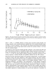
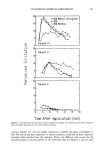
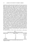
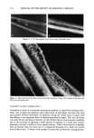
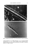
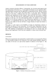
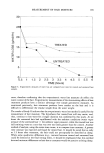
PREPARATION OF LIPOSOMES 153 and the radioactivity of the eluate was monitored. Once the eluate showed a negligible amount of tracer, a second load of the suspension was applied to the same column. Liposomes collected in the void volume were then assayed for total radioactivity. The quantities of markers recovered as well as total mass balances were compared with their counterparts where no pre-equilibration was performed. The effect of hydration temperature on the quantities of lipids incorporated into lipo- somal formulation 2 was also investigated. Liposomes were prepared by the conventional hydration method at 55 and 80øC. The quantities of lipids that participated in liposome formation at both temperatures were assessed by gel permeation chromatography. Confirmation of liposomeformation. The existence of liposomes was confirmed by viewing the lipsomal fraction eluted in the void volume under a phase-contrast microscope (Nikon Inverted Microscope, model Diaphot-TMD, Japan). When liposome formation was sparse and difficult to view, a trace quantity of 16 anthroyl oxyl palmitate, a fluorescent probe, was introduced into the suspension during the 45-min anneal period after hydration was complete. The probe is presumed to behave in a manner similar to palmitic acid and should not affect the liposome's properties since only trace quantities are used. Inclusion of the marker causes the vesicles to fluoresce when viewed under UV light. In cases where liposome formation was questionable, distribution of the radio- active marker in the column was determined. The Sephadex packing was recovered and separated into two portions. The upper portion occupied the volume from the first three centimeters of the total column height and approximated one-eighth of the entire packing material, and the lower portion represented the remainder. These packing materials were mixed with 0.2 g NaCI per 4 g Sephadex and extracted twice with a mixture of chloroform:methanol (9:1 v/v). The organic phase was collected and the solvents removed by air drying. The residue was then assayed for radioactivity. Monolayer studies Monolayer interaction studies were used to model bilayer-bilayer interactions between various lipid components. There is much precedence in the use of monolayer studies to predict interactions likely to occur in the bilayers (10). These studies were carried out to screen certain synthetic lipid mixtures to determine compositions that exhibit inter- lipid interaction potential similar to those obtainable with lipid extracts from mouse skin stratum corneum. The rationale for this approach involves the concept that if a synthetic lipid mixture displays a strength similar to that of monolayer interlipid interactions as the stratum corneum lipids, then it is highly likely that interactions between bilayers that are formed from such lipids will also be similar. Thus, compo- sitions of the synthetic lipid mixtures that show similar interaction potential as the stratum corneum extracts could then be used for the preparation of model bilayer membranes in the next phase of the study. In these experiments, the stability of monolayers formed by spreading mixtures of lipids on an aqueous subphase was investigated. This was done by monitoring changes in the surface pressure of a lipid-covered surface as a function of time using a tensiometer (Roller Smith Precision Tensiometer, Laboratory Products Inc., Boston, MA). All lipids except palmitic acid were used as obtained from suppliers. Trace surface-active impu- rities in palmitic acid were removed by overnight incubation of the lipid dissolved in
Purchased for the exclusive use of nofirst nolast (unknown)
From: SCC Media Library & Resource Center (library.scconline.org)