296 JOURNAL OF THE SOCIETY OF COSMETIC CHEMISTS (13) (14) E. Benedetti, P. Vergamini, and G. Spremolla, FT-IR analysis of single human normal and leukemic lymphocytes. Mikrochim. Acta, 1, 139-141 (1988). E. Benedetti, L. Teodori, M. L. Trinca, P. Vergamini, F. Salvati, F. Mauro, and G. Spremolla, A new approach to the study of human solid tumor cells by means of FT-IR microspectroscopy, Appl. Spectr., 44, 1276-1280 (1990). (15) M. Joy and D. M. Lewis, The use of Fourier transform infrared spectroscopy in the study of the surface chemistry of hair fibres, Int. J. Cosmet. Sci., 13, 249-361 (1991). (16) E. Benedetti, F. Papineschi, R. Consolini, and G. Spremolla, Analytical infrared spectral differences between human normal and leukaemic cells (CCL)•I, Leuk. Res., 8, 483-489 (1984). (17) E. Benedetti, M.P. Palatresi, P. Vergamini, F. Papineschi, and G. Spremolla, New possibilities of research in chronic lymphatic leukaemia by means of Fourier transform-infrared spectroscopy--II, Leuk. Res., 9, 1001-1008 (1985). (18) E. Benedetti, M.P. Palatresi, P. Vergamini, F. Papineschi, M. C. Andreucci, and G. Spremolla, Infrared characterization of nuclei isolated from normal and leukaemic (B-CCL) lymphocytes: Part III, Appl. Spectr., 40, 39-43 (1986). (19) L. C. Sperling, Hair anatomy for the clinician, J. Am. Acad. Dermatol., 25, 1-17 (1991). (20) L. C. Sperling and C. P. Samlaska, Hair anatomy as seen by light microscopy, J. Assoc. Mil. Der- matol., 15, 16-28 (1989). (21) A. IV[. Kligman, The human hair cycle, J. Invest. Dermatol., 33, 307-316 (1959). (22) E. Benedetti, B. Bottici, P. Vergamini, and G. Spremolla, Comparative Fourier transform-infrared analysis of lipids extracted from human normal and leukemic lymphocytes: Part V, Chemistry Today, 6, 57-60 (1988).

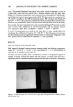
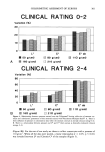
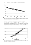
J. Soc. Cosmet. Chem., 47, 297-305 (November/December 1992) Squamometry: The assessment of xerosis by colorimetry of D-Squame adhesive discs G. E. PI•RARD, C. PI•RARD-FRANCHIMONT, D. SAINT LINGER, and A. M. KLIGMAN, Department of Dermatopathology, University of Liege, CHU du Sart Tilman, B-4000 Liege, Belgium (G.E.P., C.P.-F.), Laboratoire de Recherche Appliquge, L'Orgal, F-92117 Clichy, France (D.S.L. ), and Department of Dermatology, University of Pennsylvania, Philadelphia, PA 19104-6142 (A.M.K. ). Received April 28, 1992. Synopsis Clinical grading of the level of scaling in winter xerosis is highly subjective and crude. We have developed noninvasive objective methods to obtain more reliable information. Photographic records of epiluminescence microscopy and of cyanoacrylate skin surface biopsies enable greatly improved visualization of the scaly surface. These evaluations, however, remain semiquantitative. A real quantitative assessment is achieved by collecting corneocytes on adhesive discs (D-Squame ©) under standardized pressures, and by staining them with toluidine blue and basic fuchsin. The specimen is then subjected to colorimetry in the chroma C'mode to estimate the quantity of scales. INTRODUCTION Laymen and experts agree on what constitutes "dry skin." The surface shows scales and is rough to the touch. Dry skin, or xerosis, reflects abnormal desquamation of the horny layer in aggregates of corneocytes large enough to be seen by the naked eye as whitish scales and thin flakes (1,2). There is considerable controversy concerning the nature and origin of xerosis, a common problem in the cold winter months, especially for the elderly. Efforts to understand the etiology of dry skin and to design more effective treatments are hampered by lack of a method for quantifying the degree of scaling. Adhesive-coated slides, pressed briefly to the surface, offer a simple way to collect the clusters of corneocytes that are about to be shed from the outermost stratum corneum. Semiquantitative estimates of the size and density of scales can be made after staining. This ancient method has recently been updated by the development of D-Squame © (Cuderm Corporation, Dallas, TX). These are adhesive-coated discs of a fixed size that 297
Purchased for the exclusive use of nofirst nolast (unknown)
From: SCC Media Library & Resource Center (library.scconline.org)