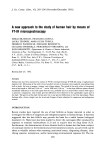
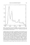
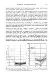
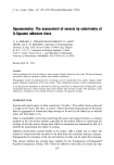
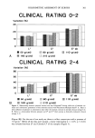
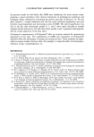
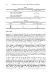
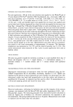
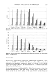
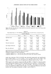
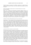
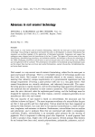
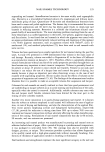
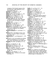
298 JOURNAL OF THE SOCIETY OF COSMETIC CHEMISTS are used in the same way. Serup and coworkers have used attenuation of optical trans- mission to measure the quantity of scales on the discs (3). We have elaborated a novel method to increase the accuracy and reproducibility of the D-Squame © technique, abetted further by utilization of cyanoacrylate surface biopsies (1,4). MATERIALS AND METHODS We collected samples of a desquamating stratum corneum from the volar forearms of adults, ages 23-56. Forty-seven had normal skin, 36 had sensitive "dry" skin, and 61 suffered from severe winter xerosis. The intensity of xerosis was estimated clinically on an analogue scale ranging from 0 to 6, based on the classification of Lukacovic et al. (5). At the same time we photographed the surface under standard conditions, using an epiluminescence camera (Dermaphot Heine, Herrsching, Germany). This method was originally designed for the examina- tion of pigmented lesions after applying oil on the surface of the skin to render the stratum corneum transparent (6,7). This procedure was modified in our study. The camera was gently applied to the surface of the skin without interposition of oil. The picture obtained then represents a standardized high magnification of the surface of the stratum corneum (Figure 1). e '• d Figure 1. Examples of epiluminescence photographs of the stratum corneum graded 0 (a), 1 (b), 3 (c), and 6 (d).

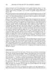
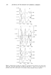
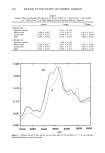
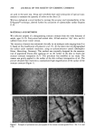
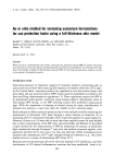
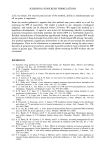
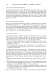
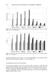
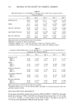
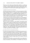
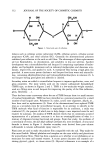
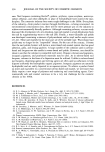
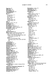
COLORIMETRIC ASSESSMENT OF XEROSIS 299 We also used terephtalate polyethylene sheets (Melinex O, ICI Plastic Division) coated with cyanoacrylate glue (Bison © Super-colle, Perfecta Chemie, Goes, Holland) to re- move surface scales, as previously described (1,4). The cyanoacrylate skin surface biop- sies (CSSB) were stained with a solution of toluidine blue and basic fuchsin (PMS: Polychrome Multiple Stain, Dermatologic Lab Supply, Bluffs, USA). The specimens were then graded (4) accoMing to the following scheme depicted in Figure 2: 0: Normal stratum corneum. 1: Hyperkeratosis of the 1st and 2nd lines and/or of the appendageal orifices. 2: Scales covering less than 30% of the plateaus. 3: Scales covering more than 30%. 4: Diffuse confluent scales. 5: Thick, uneven scales covering the entire surface, obliterating the 1st lines. We also sampled the outer horny layer by means of D-Squame © discs (Cuderm Cor- poration, Dallas, TX). These were applied with a dyanometer under standardized pres- sures of 50, 80, 110, 160, and 210 g/cm 2, respectively, to adjacent sites. These were weighted before and after sampling on a Mettier balance (Zurich, Switzerland) the accuracy of which reaches 0.1 mg. Thus the weight of the stratum corneum sample on each tape was measured as the difference in the weight before and after sampling the skin surface. The samples were then stained for one minute by dropping the staining solution PMS over the surface, followed by gentle rinsing with tap water. The stain solution and rinse water were centrifuged to insure that there was not an appreciable loss of corneo- Figure 2. Examples of CSSB graded 0 (a), 1 (b), 3 (c), and 5 (d).
Purchased for the exclusive use of nofirst nolast (unknown)
From: SCC Media Library & Resource Center (library.scconline.org)