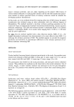
286 JOURNAL OF THE SOCIETY OF COSMETIC CHEMISTS portance of human hair in such applications is due to several important factors: it is very sensitive to the environmental conditions, its epithelial origin might represent the specific target of many carcinogens, and it contains very rapidly (anagen phase) prolif- erating cells. FT-IR spectroscopy has been extensively used in the study of biological systems in order to obtain indications (at the molecular level) on structural and chemical-physical prop- erties (7,8). Several IR approaches to the study of hair are reported in the literature, especially with respect to cosmetology (9). Because of the difficulties in obtaining hair spectra, these investigations were carried out by means of special techniques such as the diffused and the attenuated total reflectance that are known to present serious problems of reproducibility (9). Today high-resolution analytical techniques make it possible to adopt a global approach to the investigation of biological systems. FT-IR microspectroscopy, which couples the visible image to the corresponding infrared spectrum, was initially employed in the analysis of polymeric materials, especially for the study of the separation phase in polymer blends (10) and for the detection of impurities in plastic materials. Successively, the method was extended to the study of normal and leukemic single cells (11-13) and of human solid tumor cells (14). Recently the hair shaft, treated with two kinds of oxidizing agents, has been examined at different distances from the root by means of FT-IR microspectroscopy (15). Analyses of single fibers treated with hydrogen peroxide or metabisulphite solutions showed the formation of cysteic acid and cysteine-S-sulphonate, respectively further- more, this method allowed to be constructed a profile of the oxidative damage from the root to the tip of naturally weathered hair. In this investigation, we have extended the FT-IR microspectroscopy study to the entire length of the hair, examining microregions, proceeding from bulb to shaft for anagen, catagen, and telogen hair, and trying to obtain reliable and reproducible spectral data able to distinguish between hairs in the different phases of the cycle. Using this method, we can obtain information at the molecular level, both with respect to the degree of hair aging and alterations of cell differentiation due to exogenous and endogenous chemical and physical agents. EXPERIMENTAL Human hairs were plucked from the scalps of healthy male and female volunteers aged 20-50 years. Actively growing (anagen), catagen, and telogen hairs with an intact bulb were carefully selected (microscopically x 15). Full-length single hairs of normal sub- jects with the root "club" intact were subjected to no prior chemical treatments and only basic grooming, i.e., brushing, combing, and shampooing. Before analysis, all full- length hairs were washed with an aqueous solution of sodium lauryl sulphate (SDS) (10 g 1- •) for 5 minutes, thoroughly rinsed, and left to air dry. Distilled water was used for all experiments. SDS solution is a suitable means of removing any fat on the fibers, as reported by Joy et al. (15). It is important that the infrared spectra carried out on hairs before and after this treatment did not show any spectral differences even though structure modifications could not be excluded. A Spectra-Tech IR Plan optical microscope was coupled to the spectrophotometer to allow visual observation of a sample, followed by infrared analysis of the observed sites.

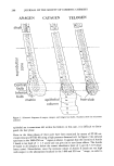
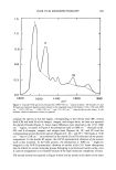
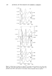
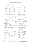
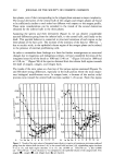
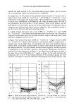
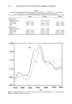
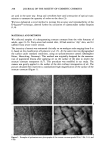
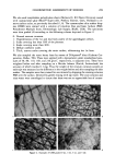
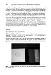
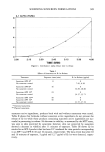
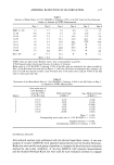
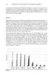
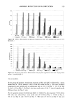
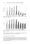
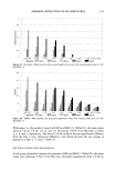
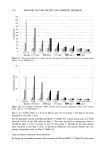
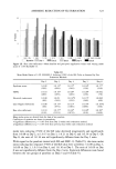
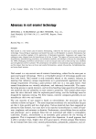
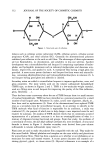
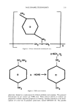
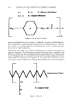
HAIR FT-IR MICROSPECTROSCOPY 287 A Perkin Elmer 1760-X spectrophotometer (MCT detector) was used for the measure- ments. Three hundred scans of 4 cm-• resolution were signal averaged. For the interpretation and band assignment of the IR spectra reference standard of a human albumin, DNA from salmon sperm and RNA from calf liver were used as --1 illustrated in previous papers (16, 17, 18 respectively). The bands at 1080 cm (mainly DNA) and at 1540 cm- • (protein) are particularly interesting since they show different features, especially in pathological samples (14). Single fibers were examined by pressing them into two diamond windows of a high- pressure diamond cell suitable for FT-IR microspectroscopy. Unlike other techniques, the microscope attachment allows the analysis of individual fibers. To obtain reliable spectra, the samples had to be suitably pressed in order to obtain optical density values as low as possible (0.7-0.9), especially for the Amide II absorption. The morphology of the sample squeezed into the high pressure diamond cell was sufficiently well main- tained, as the various regions of the hairs were found to be well defined. The spectra on thirteen-fifteen microdomaines, of the order of 100 microns in diameter, were obtained, proceeding from the bulb matrix to the shaft (for each bulb) (Figure 1). The integrated areas were calculated using the "area" command for the CDS 3-program of Perkin-Elmer computer model 7300. In Figure 2, the typical spectrum of hair is reported. Integrated areas of the bands at 1540, 1238, and 1080 cm-• were calculated for each spectrum on the basis of 1595-1475 cm-• 1300-1185 cm-• and 1137-997 --1 cm baseline points respectively, considering that in all spectra corresponding to the various phases the slope of the baselines were similar, following the trend of the background of the spectrum. The spectra of Figures 2, 3, 4, and 7 are flattened in the range considered. RESULTS AND DISCUSSION A diagram of plucked human hairs in three different stages of their cycle, as they appear at the microscopic analysis (15 X), is presented in Figure 2. The hair fiber in the diagram has been divided into three regions along its axis: bulb (inferior and central), supra- bulbar zone, and shaft. The bulb of anagen hair (matrix) is the actively growing portion, whose cells rapidly divide and move upward. It should be remembered that a plucked hair usually represents only a portion of the normal follicle, since some epithelial and all derreal components (dermal papilla and fibrous sheath) are normally left behind (19,20). This results in a "naked" hair shaft and root. The next portion in the direction of the hair shaft is the keratinization region, where the hair fiber undergoes hardening or solidification through cystine cross-linking (6). In the catagen stage the actively growing hair undergoes transformation into a dead club hair the matrix cells become separated from the connective tissue of the papilla by a column of epithelial cells (EC). The epithelial column below the club hair, although fragile, may be extracted as a clear "tail" of tissue clinging to the keratinized club (21). These features distinguish plucked catagen hairs from anagen hairs. The lowermost end of the disconnected hair shaft (hair club) becomes fully keratinized. At the beginning of the telogen phase, a remnant of the epithelial column remains as a small bud (called epithelial sac) off the base of the hair follicle. When the telogen hair is plucked, the
Purchased for the exclusive use of nofirst nolast (unknown)
From: SCC Media Library & Resource Center (library.scconline.org)