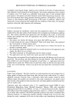
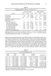
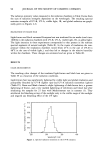
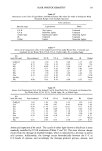
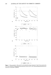
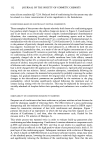
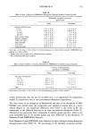
124 JOURNAL OF THE SOCIETY OF COSMETIC CHEMISTS 1.00 0.80 0.60 0.40 0.20 0.00 0 6 12 18 24 TIME (hours) Figure 2. Time course of percutaneous absorption of Padimate-O and NMPABAO in human skin. Padi- mate-O and NMPABAO were applied to skin in 15 }xl vehicle/cm 2. Padimate-O and NMPABAO were applied at chemical doses of 6.7 and 7.0 p,l/cm 2, respectively. • = Padimate-O ß = NMPABAO. Table V Absorption of Padimate-O and NMPABAO Through Human Skin Percent of applied dose absorbed PadimateoO NMPABAO Receptor fluid 0.99 + 0.26 0.73 -+ 0.15 Stratum corneum• 1.5 -+ 0.24 1.3 -+ 0.29 Viable skin 2 0.26 -+ 0.03 0.27 + 0.06 Total absorbed 2.8 -+ 0.35 2.3 -+ 0.43 24-h wash 89.6 -+ 7.3 87.8 -+ 4.0 Total recovered 92.3 - 7.2 90.1 + 4.0 Each value is the mean -+ S.E. of four determinations in each of two human skin specimens. Padimate-O and NMPABAO were applied in a lotion vehicle. • Surface layer of skin removed by cellophane-tape stripping. 2 Skin remaining after tape stripping. lized in the receptor fluid fractions ranged from 32-36 for Padimate-O to 76-92 for NMPABAO. The absorbed material remaining in the skin was essentially unmetabo- lized. As was the case in hairless guinea pig skin, NMPABAO was metabolized more extensively than Padimate-O in human skin. Greater metabolism (percent of absorbed dose) of both compounds was observed in the human skin studies. This may have occurred because saturation of metabolic enzymes in skin was prevented when absorption was reduced in the human skin experiments.

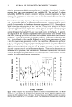
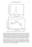
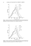
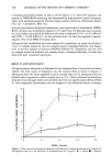
PADIMATE-O 125 Table VI Ester Hydrolysis of Padimate-O and NMPABAO in Human Skin Percent of absorbed dose metabolized Padimate-O NMPABAO Receptor fluid fraction 0-6 h 32.8 -+ 16.3 81.9 -+ 8.5 6-12 h 36.1 -+ 9.2 76.5 -+ 9.7 12-18 h 36.7 -+ 10.2 84.7 -+ 7.9 18-24 hr 32.9 -+ 6.7 92.2 -+ 3.6 Skin 0.4 -+ 0.40 0.0 -+ 0.0 Padimate-O was hydrolyzed to DMABA, and NMPABAO was hydrolyzed to NMPABA. Each value is the mean -+ S.E. of four determinations in each of three human skin specimens. The stability of NMPABAO in sunlight was investigated by exposing cosmetic formu- lations to UV light from a solar simulator. In preliminary studies, a sunscreen formu- lation spiked with NMPABAO at 7950 ppb was exposed to UV light. After a 2-min exposure (UVB dose of 0.48 mJ/cm2), only 10% of the nitrosamine was still intact (Table VII). The effect of UV irradiation on the percutaneous absorption and metabolism of NM- PABAO was determined by applying NMPABAO in the lotion vehicle to excised hairless guinea pig skin in diffusion cells and irradiating the skin with UV light (UVB dose of 5 mJ/cm2). The absorption profile of NMPABA in irradiated skin (Table VIII) was very similar to that of NMPABA in nonirradiated skin (Table III). However, UV irradiation had an important effect on the chemical species penetrating the skin. An unidentified component, presumably a photodecomposition product of NMPABAO, was found in the thin-layer chromatogram of the receptor fluid from irradiated skin. The majority of the radioactivity (88%) was found in this component (Figure 3). Only 5.9 and 6.2% of the radioactivity were identified as NMPABAO and NMPABA, respectively. The radioactivity found in the skin at 24 h was almost com- pletely decomposed by UV irradiation to the unidentified compound (2.5% was NM- PABAO). On the basis of our studies (Table VIII, Figure 3), UV irradiation of hairless guinea pig skin would reduce absorption to 0.025 ng/cm, • a 17.2-fold decrease. A sunscreen user who is exposed immediately to the sun may have reduced absorption of NMPABAO because of the instability of the compound in UV light. However, many cosmetic users regularly apply cosmetic products containing Padimate-O regardless of whether expo- Table VII Photodecomposition of NMPABAO in a Sunscreen Lotion Irradiation time (min) NMPABAO (ppb) 0.00 7950 1.00 95O 1.50 815 2.OO 775 Each value represents the mean of three repetitions for each time interval following exposure to UV light.
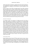
Purchased for the exclusive use of nofirst nolast (unknown)
From: SCC Media Library & Resource Center (library.scconline.org)