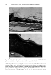
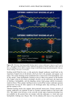
228 JOURNAL OF THE SOCIETY OF COSMETIC CHEMISTS Figure 4. (a) Damage to outer layers of corneocytes (arrows) was seen in 25% of the formulation B-treated tissue (24,000x). (b) A higher magnification view of the damaged region showing fingerprint (arrows) of lipid-like regions surrounding the outer corneocytes (128,000x). induced structural changes in epidermis with TEWL. Different surfactant treatments resulted in variable degrees of lipid and corneocyte preservation. Most of the water- washed tissue (control) displayed intact disjunctum comprising few layers (one to two) of lipid covering the outer corneocytes. In contrast, isolated regions that lacked most of

SURFACTANT-SKIN INTERACTIONS 229 b Figure 5. (a) Occasional presence ofdisjunctum in formulation C-treated tissue (TEWL 11 g/m 2 hr). Outer lipid layers (arrow) were generally featureless (24,000x). (b) Gross intracellular damage (arrowheads) and loss of intercellular lipids (arrows) can be seen in this high-magnification view of formulation C-treated tissue (128,000x).
Purchased for the exclusive use of nofirst nolast (unknown)
From: SCC Media Library & Resource Center (library.scconline.org)