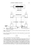
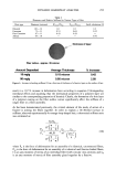
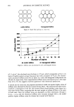
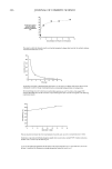
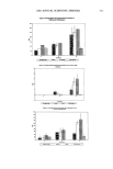
270 JOURNAL OF COSMETIC SCIENCE Though their mechanism of action is not yet fully understood, HAs are also thought to stimulate synthesis of collagen and mucopolysaccharides in the dermis (6). At use levels under 10 percent, skin care benefits are derived through a continued pattern of product usage. Continued use of products with hydroxy acid levels below 10 percent has been shown to result in a gradual reduction of fine lines and an improvement in skin texture through accelerated desquamation. At higher levels (greater than 30 percent), HAs function as chemical peeling agents that rapidly thin the stratum corneum. At these higher concentrations they achieve the same perceptible reduction in fine lines and an improvement in skin texture, though these effects are manifested more rapidly (7). One important question in the HA formulation is the relationship between the pH and topical efficacy of the formulation. The efficacy of HAs is related to the free acid concentration of the formulation--the lower pH, the higher the free acid concentration of the formulations and the more efficient the formulation (8,9). However, there is a clear inverse relationship between the pH of HA preparations and their potential for skin irritation. The lower the pH, the higher the irritation the higher the pH, going towards neutrality at 4.4, the more the irritation drops off (10). Recently emerged in the cosmetics market have been HA derivatives as substitutes for HAs in formulations. These products did not interfere with normal skin pH values, and they claim to maintain the HAs' activities (11,12). Identifying other compounds with similar effect and improved tolerance would be greatly valued. The fruit acids mixture has also been suggested as effective and safe to use in cosmetic products (11). The objective of this study was to investigate the histopathologic, morphometric, and stereologic alterations caused by the application of the cosmetic formulations containing glycolic acid, lactic acid, a fruit acids mixture, malic acid ester, or salicylic esters of lipophilic acid on the hairless mice epidermis. MATERIAL AND METHODS FORMULATIONS STUDIED The formulation consisted of 20% cetearyl alcohol/ceteareth-20, 1% squalane, 3% pro- pylene glycol, 0.15% methyl dibrome glutaronitrile, phenoxy-ethanol, and distilled water. The cream formulation was supplemented or not with 8% glycolic acid 70%, 5% lactic acid 85%, 3% fruit acids mixture, 5% malic acid ester, or 1% salicylic esters of lipophilic acid. BIOLOGICAL ASSAY Animals. Adult male hairless mouse, weight on average 30 g, were used. The animals where kept in individual cages and received a commercial ration and green food (rami), as well as water ad libitum. Treatment. One group of five animals was used as a control and the other six groups of five animals were used for application on the back area of 1 cm 2 of the cream formulation or the cream formulation containing HAs or their derivatives. The formulations were applied once a day for 15 days.

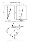
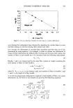
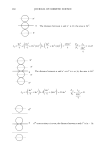
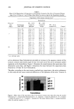
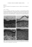
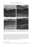
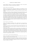
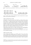
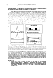
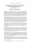
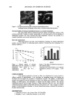
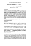
HYDROXY ACIDS IN COSMETIC FORMULATIONS 271 HISTOLOGY After the treatment periods, the hairless mice were sacrificed and skin fragments were obtained from each area and immediately immersed in a fixing solution of 85 ml of 80% alcohol, 10 ml of formalin, and 5 ml of acetic acid. The fragments were fixed for 24 hours and then dehydrated, cleared, and embedded in paraffin. Serial 6 lam-thick sections were then obtained and ten sections per block were obtained from a total of 500 sections, so that each of these ten sections would correspond to an interval of 50 sections. The sections were stained with hematoxylin and eosin. MORPHOMETRY AND KARYOMETRY For the morphometric study (analysis of the nucleus of the epithelial layers), the skin sections obtained from each experimental group were analyzed with a Hennaed light microscope equipped with a 100x immersion objective. The largest and smallest diameters of the nuclei of the basal and spinous layers of the epidermis were determined by the KS software, where the image were captured, stored, and analyzed. The following karyometric parameters were estimated: © Mean diameter: M = (D ß d) •/2 © Volume:V: 6 -• -•r'M 3 STEREOLOGY The present study was used a grid idealized by Merz (9), printed on paper, to draw the epithelial structures. The grid consists of a square that limits the test area, containing a system of points marked on a sinuous line formed by the succession of enchained semicircles, shown in Figure 1. Figure 1. The Merz grid.
Purchased for the exclusive use of nofirst nolast (unknown)
From: SCC Media Library & Resource Center (library.scconline.org)