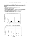
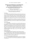
2002 ANNUAL SCIENTIFIC SEMINAR 301 References: I. Beth G. Goldstein, Practical Dermatology, 1997 46-47 2. F. Neumann, W. EIger, "The effect of a new antiandrogenic steroid, 6-Chloro-17-Hydroxy-lct, 2ct-Methylene-pregna-4, 6-Ddiene-3, 20 Dione Acetate (Cyproterone acetate) on the sebaceous glands &mice," J Invest Derre. 1966 46:56 I 3. A. Archibald, S. Shuster, "Bioassay of androgen using the rat sebaceous gland,"d. Endocr. !967 37:XXll 4. Cunliffe W. J., Shuster S., "The effect of topical cyproterone acetate on sebum secretion in patients with acne," Br. J Dermatol., 1969 81:200-201 5. Sciarra F., Toscano V., Concolino G., "Androgen: clinical applications," d. Steroid Blochem Mol Biol., 1990 37:349-362 6. James C. S., "Antiandrogen and hormonal treatment of ache," Dermatologic Clinics 1996 14:803- 811 7. C. Beylot, M. S. Doutre, "Oral contraceptives and cyproterone acetate in female acne treatment," Dermatology 1998 196:148-152 8. R.J. Scheuplein, "Mechanism &Percutaneous AbsorptionlI. Transient diffusion and the relative importance of various rates of skin penetration," J. !nvest. Dermatol. 1967 48:79-88 9. R.L. Bronaugh, R. F. Stewart, "Methods for in-vitro Percutaneous Absorption Studies VI. Preparation of the Barrier Layer," J. Pharm Sci., 1986 75:487-491 10• G. lmokawa, M. Hatton, .1. Invest. Dermatol., 1985 84:282

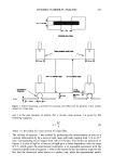
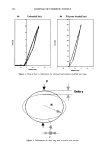
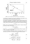
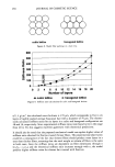
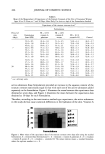
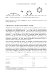
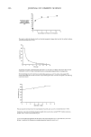
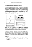
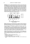
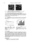
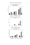
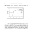
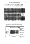
302 JOURNAL OF COSMETIC SCIENCE CHALLENGING THE SURFACTANT MONOMER SKIN PENETRATION MODEL: PENETRATION OF SODIUM DODECYL SULFATE (SDS) MICELLES INTO TUE EmDEMIS Daniel Blankschtein •, Ph.D., Peter Moore •, and Sudhakar Puvvada, Ph.D? •Department of Chemical Engineering, Massachusetts Institute of Technology, Cambridge, MA ZUnilever Home and Personal Care North America, Trumbull, CT 1. Introduction SDS dose-dependent skin irritation, in which skin irritation increases as the SDS concentration in the contacting solution increases beyond the critical micelle concentration (CMC), has been observed by several researchers (1,2). It is generally accepted that only the surfactant monomers are able to penetrate into the stratum corneum (3). However, the SDS monomer concentration is essentially constant above the CMC, and therefore, the skin irritation induced by SDS should be independent of the SDS concentration (3). Nevertheless, it has been observed that the penetration of SDS into the stratum corneum increases as the SDS concentration increases above the CMC (4,5), corresponding to the observed dose-dependent increase in skin irritafion• We propose that this apparent paradox can be resolved by demonstrating that SDS micelies are capable of penetrating into the skin. We also propose that preventing this micelle penetration should lead to a dose-independent skin irritation. 2. Materials and Methods SDS and Polyethylene Glycol (PEG, MW 8000) were obtained from Sigma Chemicals. •4C-SDS was purchased from American Radiolabeled Chemicals. Porcine skin was purchased from a local farm_ The porcine skin was exposed to a solution of SDS or of SDS+PEG for 5 hours in a diffusion cell, the skin was heat stripped, and the concentxation of SDS in the epidermis was determined using scintillation counting. 3. Results and Discussion As reported previously by other researchers (4,5), we observed that increasing the concentration of SDS in the contacting solution from 50 mM to 100 mM led to an increase in the concentration of SDS in the epidermis (C•,), see open bars in Figure 1. When SDS was mixed with 2.5wt% PEG, we observed a drop in C,•a• at all the SDS concentrations examined, and interestingly, no increase in C•-• with SDS concentration was observed (see solid bars in Figure 1). It is apparent that the addition of PEG affects the ability of SDS to penetrate into the epidermis. PEG and SDS interact to form PEG-bound SDS micelies, in which the PEG forms a corona surrounding the SDS micelies. At the SDS and PEG concentrations examined in Figure 1, only PEG-bound micelles are present in the SDS+PEG solutions, and only free SDS micelies are present in the SDS solutions (6). We hypothesized. that the lack of increase in C• with SDS concentration observed for the SDS+PEG system occurs because the free SDS micelies contribute to C,•n, while the PEG-bound SDS micelies do not. To test this hypothesis, C• was measured for 100 mM SDS with an increasing PEG concentration (see Figure 2). At low PEG concentrations, there is an excess of SDS, and the solution containes a mixture of free and PEG-bound SDS micelies. As the PEG concentration increases, the concentration of free SDS micelies decreases while the concentration of PEG-bound SDS micelles increases, until, at 1.7wt% PEG and beyond, only PEG-bound SDS micelles are present (see dotted line in Figure 2). As long as free SDS micelles are present, the SDS monomer concentration should remain constant Accordingly, a plausible explanation for the decrease in C,• observed with increasing PEG concentration is that SDS in the PEG- bound SDS micelies does not contribute to C•, A regression analysis of our results confirmed this explanation, showing that, on a per molecule basis, SDS in monomefic form contributes 4 times more to C• than SDS in free miceliar form, while SDS in PEG-bound SDS micelies does not contribute to C•. Using Dynamic Light Scattering (DLS), the hydrodynamic radii of the free and the PEG-bound SDS micelies were determined to be 20• and 25•, respectively. Researchers have reported values of aqueous pore radii in the skin to be between 10• and 28]t (7,8). We propose that the free SDS micelies are able to access these aqueous pores, while the PEG-bound SDS micelies are sterically hindered from doing so. 4. Conclusions We have found that the observed dose-dependent penetration of SDS into the epidermis is due to the contribution of miceliar SDS, thus contradicting the widely-accepted monomer penetration model. However, PEG-bound SDS micelies did not contribute to SDS penetration into the skin. Based on the
Purchased for the exclusive use of nofirst nolast (unknown)
From: SCC Media Library & Resource Center (library.scconline.org)