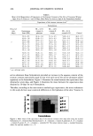
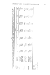
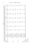
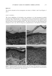
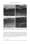
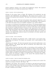
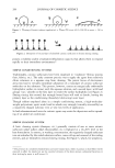
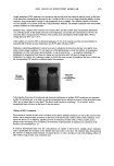
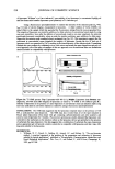
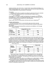
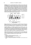
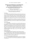
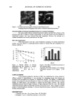
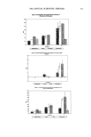
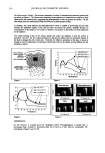
278 JOURNAL OF COSMETIC SCIENCE present significant changes in the cellular and cytoplasmatic volume. The numerical density decreased in the basal and spinous layers (Table II). GROUP V (CREAM + FRUIT ACIDS MIXTURE) Histology. In this group, shown in Figure 3B, thickness of the epithelium was also observed, with hyperkeratosis and parakeratosis. Also noticed was an increase in the thickness of the spinous layer (acantosis). The dermis was thickened in relation to group controls. Morphometry and stereology. The basal and granular layers presented alterations in the shape coefficient. The contour index was altered only in the basal layer. The eccentricity in the basal and spinous layers was modified compared to the control (Table I). An increase in the thickness of the epithelium was observed. The basal and spinous layers were thicker in relation to the control and to the cream-only group. The cellular volume was increased in the basal and spinous layers in relation to the control, and the granular layer presented a significant increase in relation to the control and to the cream-only group. There was increase in the nuclear volume in all epithelial layers in relation to the control. The cytoplasmatic volume of the basal and granular layers was increased in relation to the control. The numerical density was decreased in the basal, spinous, and granular layers (Table II). GROUP VI (CREAM + MALIC ACID ESTER) Histology. The epidermis was thickened, with a visible increase in the spinous layer. Parakeratosis was observed. The dermis was increased, with a small number of cells (Figure 3C). Morphometry and stereo/ogy. The basal and granular layers presented modifications in the shape coefficient. The contour index and the eccentricity were altered only in the granular layer (Table I). In this group we have also observed an effect on the thickness of the epithelium. The thickness of the basal and spinous layers was increased in relation to the control and to the cream-only group. The granular layer was thickened in relation to the control. The cellular volume of the spinous layer presented an increase in relation to the control. The nuclear volume was shown to be larger in the spinous layer. In this layer the cytoplas- matic volume was increased in relation to the control. The numerical density was decreased in the spinous layer (Table II). GROUP VII (CREAM + SALICYLIC ESTERS OF LIPOPHILIC ACID) Histology. A few differences were noticed in the epidermis in relation to the control. The dermis was increased, with few cells (Figure 3D). Morphometry and stereo/ogy. The eccentricity was altered in the basal and spinous layers (Table I). The thickness of the basal layer was increased. The thickness of the granular layer was decreased. The cellular and cytoplasmatic volume were increased in the granu- lar layer in relation to the control.

HYDROXY ACIDS IN COSMETIC FORMULATIONS 279 DISCUSSION In this study, according to the methodology employed, we had the opportunity to evaluate and compare the effects of several HAs and HA esters. The HA esters have pH close to skin pH, a pH of 4.5-5.0, and thus they present a low irritant potential and, like the HAs, their application on the skin can cause both epidermal and dermal effects (6). Among the studied substances, only the salicylic esters mixture did not increase the epithelium thickness significantly, probably because its pH and molecular mass are higher than other studied substances. The other actives had a significant advantage over the vehicle in improving the epidermal thickness, probably due to cutaneous hydration. This result is consistent with data reported by other authors (6), where the treatment with glycolic acid, lactic acid, and citric acid caused an increase in skin thickness, with an increase in epidermal thickness. Glycolic acid, lactic acid, the fruit acids mixture, and malic acid provoked the hydration effect by intra- and extracellular edema. This effect is very interesting because it can improve cutaneous hydration, and it is not limited to the upper cell layers but is also present in the deeper ones. The application of the cream and the cream plus glycolic acid, lactic acid, and fruit mixture formulations caused an increase in the cellular volume of the basal layer and of the spinous layer. In the presence of the malic acid ester an increase in cellular volume was observed only in the spinous layer. The increase in cellular volume was at the expense of nuclear volume and/or an increase in cytoplasmatic volume (intracellular hydration). No inflammation was evident. The increased nuclear volume is probably related to the increased cellular synthesis activity and intense mitotic activity (13). Thus the active substances provoked an increased nuclear volume that may suggest that the nuclei are in intensive activity and that as a result a cellular function stimulus is occurring. The presence of glycolic acid and lactic acid in the cream formulation caused an increase in the nuclear volume of the basal layer, statistically different from the other groups. This result coincided with that of Smith (14), who studied the effects of alpha hydroxy acids (AHAs: glycolic acid, lactic acid, malic acid), and demonstrated that the glycolic acid and lactic acid were more effective in cellular renewal stimulation. Among the active substances studied, only the glycolic acid and the lactic acid caused a significant increase in the thickness of the stratum corneum, which indicates that this layer is in the process of exfoliation. Van Scott and Yu (4) demonstrated that AHAs in topical application reduce the thick- ness of hyperkeratotic stratum corneum, decreasing the corneocyte cohesion. After treat- ment the stratum corneum is less strongly linked to the interior part of the epidermis. Multiple mechanisms may be involved. Low concentrations of AHAs diminish corneo- cyte cohesion (2). The effect occurs at the lower levels of the stratum corneum, and it may involve a dynamic process operative at a particular step of keratinization, like the modification of the ionic bonding. Another mechanism involved is enzymatic inhibition, induced by AHAs, of the reactions of sulphate transferase, phosphotransferase, and kinases, leading to fewer electronegative sulphate and phosphate groups on the outer wall of corneocytes and resulting in the diminishing of the cohesion forces (1).
Purchased for the exclusive use of nofirst nolast (unknown)
From: SCC Media Library & Resource Center (library.scconline.org)