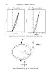
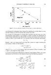
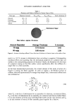
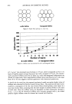
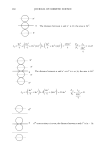
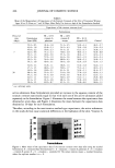
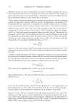
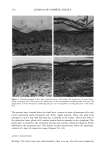
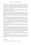
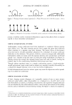
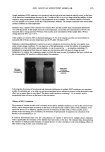
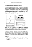
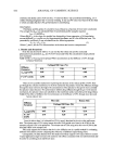
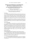
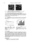
HYDROXY ACIDS IN COSMETIC FORMULATIONS 271 HISTOLOGY After the treatment periods, the hairless mice were sacrificed and skin fragments were obtained from each area and immediately immersed in a fixing solution of 85 ml of 80% alcohol, 10 ml of formalin, and 5 ml of acetic acid. The fragments were fixed for 24 hours and then dehydrated, cleared, and embedded in paraffin. Serial 6 lam-thick sections were then obtained and ten sections per block were obtained from a total of 500 sections, so that each of these ten sections would correspond to an interval of 50 sections. The sections were stained with hematoxylin and eosin. MORPHOMETRY AND KARYOMETRY For the morphometric study (analysis of the nucleus of the epithelial layers), the skin sections obtained from each experimental group were analyzed with a Hennaed light microscope equipped with a 100x immersion objective. The largest and smallest diameters of the nuclei of the basal and spinous layers of the epidermis were determined by the KS software, where the image were captured, stored, and analyzed. The following karyometric parameters were estimated: © Mean diameter: M = (D ß d) •/2 © Volume:V: 6 -• -•r'M 3 STEREOLOGY The present study was used a grid idealized by Merz (9), printed on paper, to draw the epithelial structures. The grid consists of a square that limits the test area, containing a system of points marked on a sinuous line formed by the succession of enchained semicircles, shown in Figure 1. Figure 1. The Merz grid.

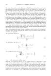
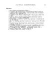
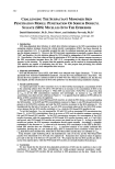
272 JOURNAL OF COSMETIC SCIENCE The Merz grid can be used to count points on a given histologic structure and also to count intersections between two contiguous structures, by considering the number of points that fall on the structure under study in the former case and the number of times that neighboring surfaces cut the curved line in the latter. Thus, in order to obtain the thickness, the numerical nuclear density, and the cytoplasm volume, we used point counting (2000 per animal, corresponding to the product of 20 microscope fields per 100 points on the grid) or the number of intersections, according to the requirements of the stereologic equation with respect to the parameter studied. Numerical nuclear density (Nvn). The area of the epithelium within the test system was evaluated by counting the points that fall on it, and the epithelial volume was propor- tional to it. The nuclei inside the standard square were then counted. The total area of the square is 50.625 pm 2 in two fields per section, for a total of 20 fields per block, and this permitted us to obtain the number of nuclear sections of the area (Nav). The number of nuclei per unit volume (numerical nuclear density, Nvn) was calculated using the Abercrombie (10) correlation modified by Elias et al. (11): Nay Nvn - D+T where D is the mean nuclear diameter previously estimated by karyometry and t is the thickness of the section (6 •m). The result obtained corresponds to the number of nuclei per mm 3. Cytoplasmic volume and epithelial cell volume. Cytoplasmic volume (Cct) was estimated from the previously determined nuclear volume and the corrected nucleus/cytoplasm ration. In turn, the sum of the mean nuclear and cytoplasmic volumes provides the estimated value of the epithelial cell. The cytoplasmic volume is given by the ratio: Vn Vct= corrected n / c The volume of the epithelial cell, in turn, is given by the equation: V cel= Vn +Vct Mean epithelial thickness. The mean epithelial thickness was estimated by the formula of Weibel (12,13): P'L 2 (IK +Ict) where P is the number of points that fell on the epithelium, L is the length of the test line, and IK and Ict are the numbers of intersections of the test line with the epithelium- keratin interface and the epithelium-connective tissue interface, respectively. Statistical analysis. Data were analyzed statistically using nonparametric tests such as the Friedman test. Data analysis and the mathematical calculations involved in the stereo- logic studies were performed using several programs elaborated in the Department of Stomatology, Dental School of Ribeirgo Preto, USP, by Profs. Geraldo Maia Campos and Miguel Angel Sala Di Matteo using Advanced Basic language.
Purchased for the exclusive use of nofirst nolast (unknown)
From: SCC Media Library & Resource Center (library.scconline.org)