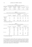
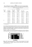
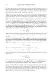
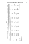
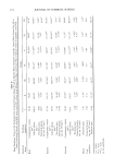
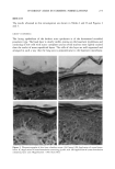
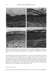
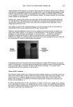
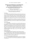
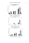
HYDROXY ACIDS IN COSMETIC FORMULATIONS 277 keratinized and there weren't nuclei in the horny layer, and an increase of the spinous layer could be observed. There was an apparent increase of the dermal thickness as compared with the control, with the presence of few cells (Figure 2B), with can suggest a new dermal collagen synthesis. Morphometry amt stereo/ogy. The shape coefficient showed significant alterations in all epithelial layers analyzed (Table I). The spinous layer presented a significant increase in thickness in relation to the control. The basal cells and their nuclei and cytoplasm were more voluminous. In the spinous layer an increase in the cellular volume could also be observed. The nuclear and cytoplasmatic volume was increased. The nuclear volume was increased in the granular layer. The numerical density was shown to be decreased in the basal and spinous layer (Table II). GROUP III (CREAM + GLYCOLIC ACID) Histology. The thickness of the epidermis was greatly increased. There was hyperkeratosis and parakeratosis, that is to say, the nucleus was gone in normal stratum corneum cells but persisted in incompletely keratinized cells. It is believed that such cells undergo differentiation too rapidly for destruction to be completed. The spinous layer was also shown to be thicker, with three to four cellular layers. The dermis was thickened and homogeneous (Figure 2C). Morphometry and stereo/ogy. The shape coefficient was changed in all the epithelial layers. The contour index and the eccentricity were altered in the basal and granular layers (Table I). The epidermis was thicker, with an increase in the thickness of the basal and spinous layers. The cellular volume in the basal and spinous layers was increased in relation to the control and in relation to the cream only. This increase in the cellular volume was connected with the increase in the nuclear and cytoplasmatic volume. An increase in the nuclear volume in the granular layer was observed. The numerical density in the basal and spinous layers was decreased in relation to the control (Table II). GROUP IV (CREAM + LACTIC ACID) Histology. The epidermis was found to have changes characterized by thickening, hy- perkeratosis, and parakeratosis. Hypergranulosis and acantosis (the spinous layer pre- sented more than four layers of cells) were observed. Edema was evident. The dermis was visibly thickened, hydrated, and hypercellular (Figure 2D). Morphometry and stereology. The shape coefficient was altered in relation to the control in all epithelial layers. Only the spinous layer did not present significant changes in the contour index. The eccentricity presented alterations only in the basal layer (Table I). This group also presented a significant increase in the thickness of the epithelium. The basal and spinous layers were thickened, and the thickness of the granular layer didn't present a significant difference in relation to the control. An increase in the cellular volume of the basal and spinous layers was observed. The nuclear volume of the all layers was increased in relation to the control. The cytoplasmatic volume of the basal and spinous layers was also increased in relation to the control. The granular layer didn't

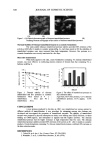
278 JOURNAL OF COSMETIC SCIENCE present significant changes in the cellular and cytoplasmatic volume. The numerical density decreased in the basal and spinous layers (Table II). GROUP V (CREAM + FRUIT ACIDS MIXTURE) Histology. In this group, shown in Figure 3B, thickness of the epithelium was also observed, with hyperkeratosis and parakeratosis. Also noticed was an increase in the thickness of the spinous layer (acantosis). The dermis was thickened in relation to group controls. Morphometry and stereology. The basal and granular layers presented alterations in the shape coefficient. The contour index was altered only in the basal layer. The eccentricity in the basal and spinous layers was modified compared to the control (Table I). An increase in the thickness of the epithelium was observed. The basal and spinous layers were thicker in relation to the control and to the cream-only group. The cellular volume was increased in the basal and spinous layers in relation to the control, and the granular layer presented a significant increase in relation to the control and to the cream-only group. There was increase in the nuclear volume in all epithelial layers in relation to the control. The cytoplasmatic volume of the basal and granular layers was increased in relation to the control. The numerical density was decreased in the basal, spinous, and granular layers (Table II). GROUP VI (CREAM + MALIC ACID ESTER) Histology. The epidermis was thickened, with a visible increase in the spinous layer. Parakeratosis was observed. The dermis was increased, with a small number of cells (Figure 3C). Morphometry and stereo/ogy. The basal and granular layers presented modifications in the shape coefficient. The contour index and the eccentricity were altered only in the granular layer (Table I). In this group we have also observed an effect on the thickness of the epithelium. The thickness of the basal and spinous layers was increased in relation to the control and to the cream-only group. The granular layer was thickened in relation to the control. The cellular volume of the spinous layer presented an increase in relation to the control. The nuclear volume was shown to be larger in the spinous layer. In this layer the cytoplas- matic volume was increased in relation to the control. The numerical density was decreased in the spinous layer (Table II). GROUP VII (CREAM + SALICYLIC ESTERS OF LIPOPHILIC ACID) Histology. A few differences were noticed in the epidermis in relation to the control. The dermis was increased, with few cells (Figure 3D). Morphometry and stereo/ogy. The eccentricity was altered in the basal and spinous layers (Table I). The thickness of the basal layer was increased. The thickness of the granular layer was decreased. The cellular and cytoplasmatic volume were increased in the granu- lar layer in relation to the control.
Purchased for the exclusive use of nofirst nolast (unknown)
From: SCC Media Library & Resource Center (library.scconline.org)