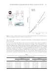
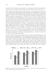
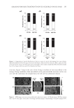
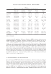
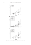
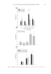
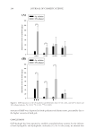
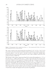
ENVIRONMENTAL PARAMETERS ON SWEAT GLAND ACTIVITY 253 temperature of 45°C and 35% RH. In all cases, except the cold acclimatization proce- dure, subjects fi rst spent 30 min in the environmental room at 27°C and 50% RH. Drinking a glass of hot water (76°C) at t = 20 min in the sauna chamber resulted in a sharp increase in the fi ring output of the sweat glands—an increase ranging from 20% to 29% for Subject 1 in the left and right axillae, respectively, and 58% and 91% for Subject 2 in the left and right axillae, respectively. As a control, we had subjects also consume a glass of room-temperature (RT) water—measured at 22°C—also at t = 20 min in the sauna chamber. In this case, there appears to be a very slight increase in sweat activity, although in most cases it falls within the limits of the standard deviation. We also conducted a clinical protocol, which incorporated physical activity into the acclima- tization period in the environmental room. The subject spent 15 min in the environ- mental room under the same conditions as in all other tests followed by a 15-min period of physical exertion on a stationary exercise bicycle (20 mph, 86 rpm, level 3). In this case, physical activity resulted in a similar increase in perspiration rate as with consump- tion of hot water. In the fi nal test listed in Table II, both subjects were acclimatized in a cold room at 2°C and 80% RH for 30 min prior to immediate entry into the sauna chamber, thereby replacing the 30-min equilibration period in the environmental room at 27°C and 50% RH. As a result, the rate of perspiration decreased markedly by 70% and 75% in the left and right axillae, respectively, for Subject 1 and 58% and 53% in the left and right axillae, respectively, for Subject 2. Such experiments offer ideas for alternative equilibration techniques when testing antiperspirant actives. For example, we might be able to reduce the amount of time spent during the acclimatization phase of the study by incorporating physical activity or hot water consumption into the test protocol. Likewise, we must also consider the subject’s environment prior to the clinical test. If the subject spends time in a cold environment prior to entry into the environ- mental room or sauna, this will certainly infl uence the outcome of the results. In fact, the inspiration for conducting the cold acclimatization test came from such conditions. When developing the test protocol, we noted that subjects who spent most of their day in a cold environment had much lower perspiration rates than on days when their cli- matic conditions were better controlled. Essentially, when subjects commented that they had a chill because their offi ce space was unusually cool on a particular day, we observed a reduction in gravimetric output. IR THERMAL IMAGING IR thermography is an imaging science concerned with the measure of emitted IR radia- tion from objects in the electromagnetic spectrum range of 9,000–14,000 nm (9–14 μm). Thermal imaging cameras are used for a variety of different temperature-related imaging solutions including medical imaging, military applications (night vision), manufactur- ing situations, and research investigations. It provides a quick approach for acquiring accurate temperature measurements in the form of an image. It is a nondestructive tech- nique that allows us to resolve the temperature distribution of objects within an image. Each image contains a two-dimensional grid (x and y), representing spatial coordinates (just like a normal digital photograph) with z information (temperature) plotted in the form of a color distribution scale. Therefore, each pixel in the image contains temperature data plotted using a distribution of various shades of colors ranging between green and white.

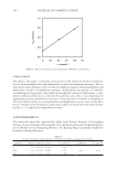
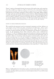
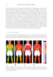
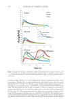
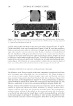
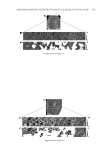
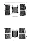
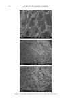
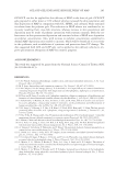
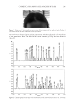
JOURNAL OF COSMETIC SCIENCE 254 Figure 4 contains a series of images we obtained for one of the subjects upon entry into the environmental room, then after 30 min in the room, followed by a period of time spent in the sauna chamber. In Figure 4A, the overall surface temperature of the skin is lower than in Figures 4B–4D. This phenomenon occurs because the subject was acclima- tized in an offi ce for several hours beforehand at a lower temperature than the environ- mental room. After 30 min in the environmental room (Figure 4B), the surface temperature of the subject begins to increase, especially in the rostral (facial) region and the superior torso. After 10 min in the sauna (Figure 4C), peak temperature is reached at the rostral region, superior torso, and the lower limbs. The subject begins to sweat pro- fusely after 10 min in the sauna, resulting in an overall cooling effect over all surfaces of the integument as shown in Figure 4D. Subsequent images were obtained at 30, 40, and 50 min, which showed similar behavior as that observed at 20 min. In summary, these data show that the 30-min acclimatization in the environmental room followed by the fi rst 10 min in the sauna chamber is suffi cient to bring the subject to a state of profuse sweating, which can be monitored for the last 20 min of the skin replica test. (A total time of 30 min was spent in the sauna chamber during the skin replica test.) FLUX DENSITY MEASUREMENTS As previously mentioned, IR imaging was used to view components of the subject’s ho- meostatic response to rising internal temperatures, including changes in radiative dissi- pation and subsequent sudorifi c cooling. On occassion, prior to testing, the subject Figure 4. (A) IR images collected upon entry into the environmental room, (B) after 30 min acclimatization in the environmental room, (C) after 10 min, and (D) 20 min in the sauna. The scale for all the images is provided on the right and ranges from 28.1°C to 37.6°C. Precise measurements of temperature can be made at every pixel in the image.
Purchased for the exclusive use of nofirst nolast (unknown)
From: SCC Media Library & Resource Center (library.scconline.org)