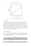
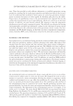
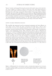
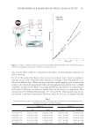
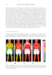
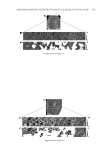
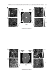
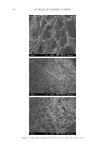
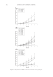
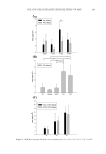
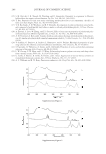
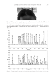
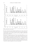
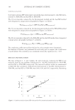
ALKALINE PEROXIDE TREATMENT INDUCES ACQUIRED UNRULY HAIR 263 Pro software (WaveMetrics, Inc., Lake Oswego, Oregon) was used to calculate the Young’s modulus from the force curve (18). AFM measurements were performed 10 times with both straight and curly hairs, and representative data were used for subsequent analyses. RESULTS AND DISCUSSION CHANGES OF APPEARANCE IN UNTREATED AND BLEACHED HAIRS Straight hairs and curly hairs were bleached and their change in shape was observed (Figures 1A and B). In the case of bleached straight hairs, changes in the length or width of the hairs were not observed, and most straight hairs remained unaltered (Figure 1A). On the other hand, in the case of bleached curly hairs, it was observed that the ROC be- came smaller than the ROC of corresponding untreated curly hairs (Figure 1B). The ratio of measured ROC in bleached curly hairs vs. corresponding untreated hairs was 0.94 ± 0.09 (mean ± S.D.), clearly demonstrating that bleach treatments decrease the ROC. COMPARISON WITH THE SPATIAL DISTRIBUTION OF TWO TYPES OF CORTICAL CELLS The differences in the distribution of inner components of straight and curly hairs were investigated by AFM measurement. Cross sections of virgin straight, bleached straight, virgin curly, and bleached curly hairs were scanned, using height and phase measurements commonly employed in topography observation. The measurements were scanned se- quentially, starting at the cuticle and proceeding through the other end, with a scan area of 10 × 10 μm. A representative AFM image of virgin straight hair is shown in Figure 2 (for other hair types, see Supplementary Figures 1–3). Height, phase, and processed phase images are shown in Figures 2A–C, respectively. Measurements were performed in water because this condition made it possible to distinguish the cell membrane complex from the intermacrofi brillar matrix (14). For reference, Figure 2D contains a zoomed image of a 5 × 5 μm region of the image in Figure 2B. Phase images painted in white by an image processor correspond to the distribution of the paracortex-like structure the remaining region is orthocortex-like structure (Figure 2C). Furthermore, the distributions of the two types of cortical cells (black: orthocortex-like structure white: paracortex-like structure) were estimated, which is shown in Figure 3. In curly hairs, cortical cells on the concave side Figure 1. Changes of appearance in untreated and bleached hairs. (A) Shapes of straight hairs and (B) shapes of curly hairs each sample was N = 20. A picture was taken of each hair sample, and the outline of the hair was traced in black to clarify the observation of the change in hair shape. Results were compiled from 20 independent hair samples.

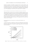
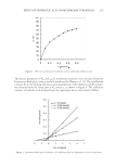
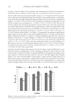
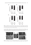
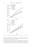
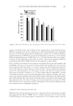
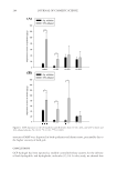
JOURNAL OF COSMETIC SCIENCE 264 could be distinguished from those on the convex side in the curly hair (Figures 3C and D). On the other hand, in the case of straight hairs (Figures 3A and B), it was not possible to defi ne concave and convex sides for convenience, straight hairs were divided into left and right sides instead of concave and convex sides. To date, it has been commonly believed that the distribution of the paracortex-like structure on the concave side was related to curliness. If this biased distribution were solely responsible for curliness, however, it would be diffi cult to explain why straight hairs (Figures 3A and B) also exh ibited biased distri- bution of the two types of cortical cells. Conversely, not all curly hairs had biased distri- bution (Figure 3D). Therefore, it was not possible to defi nitively state that the shapes of hairs were solely infl uenced by the distributions of the two types of cortical cells. COMPARISON WITH THE YOUNG’S MODULUS IN STRAIGHT AND CURLY HAIRS The differences in the Young’s modulus of the inner components of straight and curly hairs were investigated, again using AFM force curve measurement. The Young’s modulus in cross sections of virgin straight, bleached straight, virgin curly, and bleached curly hairs were measured. Representative data from bleached curly hairs is shown in Figure 4 (for other hair types, see the Supplementary Figures4–6). It was noteworthy that a difference existed between the moduli of the concave and convex sides of curly hairs (Figure 4C) this difference could hardly be observed by conventional AFM phase imaging (Figure 4B). Young’s modulus of macrofi brils was analyzed because of previous reports that the mac- rofi brils are signifi cantly infl uenced by bleach treatment (15). Young’s modulus of mac- rofi brils in the four types of hair was measured at 20 random points the average Young’s modulus is shown in Figure 5. Macrofi brils on the concave and convex side of curly hairs, or on the left and right sides for straight hairs, could be distinguished from each other in the same manner as in Figure 3. In addition to those sections, macrofi brils on the orthocortex-like structure could be distinguished from those on the paracortex-like Figure 2. AFM images of a cross section of virgin straight hair in water. (A) Height image. (B) Phase image. (C) Phase image, with the paracortex-like structure painted in white. (D) Phase image containing a magnifi ed 5 × 5 μm section of the image in Figure 2B.
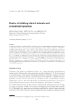
Purchased for the exclusive use of nofirst nolast (unknown)
From: SCC Media Library & Resource Center (library.scconline.org)