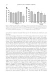
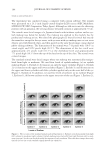
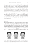
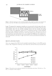
J. Cosmet. Sci., 66, 145–159 (May/June 2015) 145 Inhibitory effects of pomegranate concentrated solution on the activities of hyaluronidase, tyrosinase, and metalloproteinase SU JIN KANG, BEOM RAK CHOI, SEUNG HEE KIM, HAE YEON YI, HYE RIM PARK, SOO JIN PARK, CHANG HYUN SONG, JI HA PARK, YOUNG JOON LEE, and SAE KWANG KU, The Medical Research Center for Globalization of Herbal Medicine, Daegu Haany University, Gyeongsan (S.J.K., S.J.P., C.H.S., Y.J.L., S.K.K.), Department of Preventive Medicine, College of Korean Medicine, Deagu Haany University, Gyeongsan (S.J.K., Y.J.L.), Research Institute, Health-Love Co., Ltd, Anyang (B.R.C., S.H.K., H.Y.Y., H.R.P.), Department of Histology and Anatomy, College of Korean Medicine, Daegu Haany University, Gyeongsan (S.J.P., C.H.S., S.K.K.), and Department of Herbology, College of Korean Medicine, Daegu Haany University, Gyeongsan, (J.H.P.), Republic of Korea. Accepted for publication March 15, 2015. Synopsis Botanical antioxidants have attracted much attention as useful preventatives of skin damage. Pomegranate is consumed throughout the world for its benefi cial health effects, including its antioxidant and anti-infl amma- tory activities. We investigated whether pomegranate concentrated solution (PCS) could serve as a potential functional cos- metic ingredient that exerts a skin-whitening effect and antiwrinkle activity. To investigate the moisturizing effect of PCS, hyaluronidase activity was examined in human keratinocytes (HaCaT). Elastase and procollagenase activities were assessed in normal human primary dermal fi broblast- neonatal (HDF-N) cells to determine their antiwrinkle effects. Metalloproteinase 1 (MMP-1) activity was also assessed following ultraviolet A (UVA) irradiation. Whitening effects were measured by a tyrosinase inhibition assay and melanin formation test in mouse melanocytes (Melan-a). In addition, histopathological analysis was performed to determine the number of microfolds formed on the epithelial surface, mean epithe- lial thickness, mean number of infl ammatory cells infi ltrating the dermis, and collagen fi ber-occupied regions within the dermis. Hyaluronan synthesis was signifi cantly increased by PCS in HaCaT cells, while procollagenase and elastase activities were decreased in HDF-N cells. A signifi cant decrease in UVA-induced MMP-1 activity was also observed in PCS-treated HDF-N cells, compared with UVA-exposed cells. PCS effectively reduced melanin Address all correspondence to Sae Kwang Ku at gucci200@hanmail.net and Young Joon Lee at gksxntk@ dhu.ac.kr. Su Jin Kang and Beom Rak Choi contributed equally to this work.

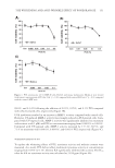
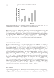
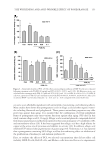
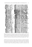
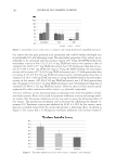
JOURNAL OF COSMETIC SCIENCE 146 production and mushroom tyrosinase activity in Melan-a cells. Moreover, UVB-induced histopathological dermal sclerosis and infl ammatory signs were signifi cantly attenuated in PCS-administered mice compared with UVB-exposed mice. Conclusions: Our results suggest that PCS prevents signs of aging, including those related to photoaging. These effects are associated with enhanced hyaluronan synthesis, as well as suppressed elastase, collagenase, MMP-1, and tyrosinase activities and melanin production. UVB-induced photoaging, such as histopatho- logical dermal sclerosis and infl ammatory signs, were effectively reduced on the addition of PCS. These re- sults also suggest that skin aging can be prevented and reduced by the antioxidant effects of PCS. INTRODUCTION Skin prevents the invasion of harmful components, serving as a protective barrier for the body. However, it is exposed to ionizing radiation, chemicals, toxins, and oxidative stressors from both endogenous and exogenous sources (1) these factors cause a loss of structural integrity and physiological function, ultimately leading to skin damage. Specifi cally, exposure of skin to ultraviolet (UV) radiation (UVA and UVB) is considered an important cause of skin aging. Consequently, skin can produce visible signs, such as fi ne wrinkles, irregular dryness, dyspigmentation, sallowness, deep furrows or severe atrophy, dehydration, skin roughness, telangiectasia, and premalignant lesions (2). UV irradiation can also induce the expression of matrix metalloproteinases (MMPs). MMPs, a large family of zinc-dependent endoproteases, have the ability to degrade all extracellular matrix (ECM) proteins (3). ECM degradation is exacerbated by aging and radiation, demonstrating a decrease in dermal collagen and an increase in MMP-1, which cleaves interstitial collagen (4). Furthermore, collagenase activity has been shown to be inhibited by tissue inhibitor of metalloproteases (TIMP) (5). Functional cosmetics have been used to prevent skin alterations, exerting potent antiag- ing properties (i.e., whitening, antiwrinkle, and moisturizing effects) (6). In particular, functional ingredients from natural sources represent very attractive materials due to their reactive oxygen species (ROS) scavenging potential and inhibition of the UV-induced signal transduction pathway (7,8). Thus, antioxidant substances may represent a promis- ing strategy for the prevention of skin aging. Pomegranates are consumed throughout the world for their benefi cial health effects. The pomegranate is comprised mostly of ellagic acid and other organic materials including fl avonoids and polyphenols (9–11). The pomegranate contains more antioxidants than both red wine and green tea. These characteristics provide protection against heart disease and cancer (12–17). In addition, the pomegranate possesses antiproliferative (18), anti- infl ammatory (19), and antitumorigenic functions (20). This evidence prompted us to examine the protective effects of pomegranate concentrated solution (PCS) on skin aging in vitro using human keratinocytes (HaCaT), mouse mela- nocytes (melanocytes), and normal human primary dermal fi broblast-neonatal cells (HDF-N fi broblasts). The antiwrinkle effects of PCS were determined through changes in hyaluronan synthesis in HaCaT cells. MMP-1 activity and procollagen synthesis were also examined in HDF-N cells due to the close correlations between wrinkle formation and loss of elasticity, collagenase, and MMP-1. Whitening effects were evaluated by a tyrosinase inhibition assay, and melanin formation was measured in Melan-a cells. In ad- dition, histopathological analysis on dorsal back skin tissue from UV-exposed mice was performed to explore the anti-photoaging effect of PCS in vivo.
Purchased for the exclusive use of nofirst nolast (unknown)
From: SCC Media Library & Resource Center (library.scconline.org)